
現在、価格および在庫状況を閲覧できません。
おすすめの製品


由来生物
mouse
品質水準
抗体製品の状態
purified immunoglobulin
抗体製品タイプ
primary antibodies
クローン
PG-4, monoclonal
交差性
human, chicken, shark
包装
antibody small pack of 25 μL
テクニック
ELISA: suitable
immunohistochemistry: suitable (paraffin)
western blot: suitable
アイソタイプ
IgMκ
輸送温度
ambient
ターゲットの翻訳後修飾
unmodified
遺伝子情報
human ... FAM20B(9917)
詳細
Proteoglycans (PG), present in all tissues, are one of the best-studied classes of matrix molecules. Over 50 members of PG have been characterized and studied for their biological significance. PG consist of a protein core to which glycosaminoglycan chains are covalently linked. Glycosaminoglycans (GAGs) are natural heteropolysaccharides that are present in every mammalian tissue. They are composed of repeating disaccharide units that consist of either sulfated or non-sulfated monosaccharides. Their molecular size and the sulfation type vary based on tissue type. The major types of GAGs found in mammalian tissues are: hyaluronan, chondroitin sulfate, and dermatan sulfate, heparin and heparan sulfate, and keratan sulfate. GAG chains are covalently bound to serine residues of the PG protein core via a tetrasaccharide linkage, consisting of xylose, two galactose residues and glucuronic acid. This antibody, clone PG-4, recognizes antigen that is associated with extracellular matrices and surfaces of cultured human dermal fibroblasts and the surfaces of cultured human adult keratinocytes. This antibody recognize native epitopes located within glycosaminoglycan chains and is detects both chondroitin sulfate (Versican) and dermatan sulfate (Decorin and Biglycan) proteoglycans. Treatment of PG with chondroitinases is reported to abolish its immunoreactivity for all antigens. (Ref.: Afratis, N et al. (2012). FEBS Journal 279 (7); 1177-1197; Sorrell, JM et al. (1999). Histochem. J. 31 (8); 549-558).
特異性
Clone PG4 detects glycosaminoglycan in skin cells.
免疫原
Crude membrane fractions from adult human dermal fibroblasts from breast skin.
アプリケーション
Research Category
細胞骨格
細胞骨格
Anti-Glycosaminoglycan, skin specific, clone PG-4, Cat. No. MABT819, is a mouse monoclonal antibody that detects glycosaminoglycan and has been tested for use in ELISA, Immunohistochemistry (Paraffin), and Western Blotting.
Western Blotting Analysis: A representative lot detected Glycosaminoglycan in Western Blotting applications (Sorrell, J.M., et. al. (1999). Histochem J. 31(8):549-58).
ELISA Analysis: A representative lot detected Glycosaminoglycan in ELISA applications (Sorrell, J.M., et. al. (1999). Histochem J. 31(8):549-58).
Immunohistochemistry Analysis: A representative lot detected Glycosaminoglycan in Immunohistochemistry applications (Sorrell, J.M., et. al. (1999). Histochem J. 31(8):549-58).
品質
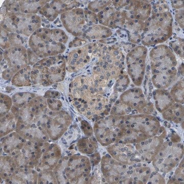
Evaluated by Immunohistochemistry (Paraffin) in human skin tissue.
Immunohistochemistry Analysis: A 1:250 dilution of this antibody detected Glycosaminoglycan in human skin tissue.
Immunohistochemistry Analysis: A 1:250 dilution of this antibody detected Glycosaminoglycan in human skin tissue.
ターゲットの説明
~60 kDa observed. Uncharacterized bands may be observed in some lysate(s).
物理的形状
Protein G purified
Format: Purified
Purified mouse monoclonal antibody IgM in buffer containing 0.1 M Tris-Glycine (pH 7.4), 150 mM NaCl with 0.05% sodium azide.
保管および安定性
Stable for 1 year at 2-8°C from date of receipt.
その他情報
Concentration: Please refer to lot specific datasheet.
免責事項
Unless otherwise stated in our catalog or other company documentation accompanying the product(s), our products are intended for research use only and are not to be used for any other purpose, which includes but is not limited to, unauthorized commercial uses, in vitro diagnostic uses, ex vivo or in vivo therapeutic uses or any type of consumption or application to humans or animals.
適切な製品が見つかりませんか。
製品選択ツール.をお試しください
保管分類コード
12 - Non Combustible Liquids
WGK
WGK 1
引火点(°F)
does not flash
引火点(℃)
does not flash
適用法令
試験研究用途を考慮した関連法令を主に挙げております。化学物質以外については、一部の情報のみ提供しています。 製品を安全かつ合法的に使用することは、使用者の義務です。最新情報により修正される場合があります。WEBの反映には時間を要することがあるため、適宜SDSをご参照ください。
Jan Code
MABT819-25UL:
MABT819:
試験成績書(COA)
製品のロット番号・バッチ番号を入力して、試験成績書(COA) を検索できます。ロット番号・バッチ番号は、製品ラベルに「Lot」または「Batch」に続いて記載されています。
Kazutoyo Terada et al.
Journal of cellular physiology, 239(2), e31174-e31174 (2023-12-18)
The Dja2 knockout (Dja2-/- ) mice had respiratory distress, and >60% died within 2 days after birth. The surviving adult Dja2-/- mice were infertile and the lungs of Dja2-/- mice showed several abnormalities, including the processing defect of prosurfactant protein
Mahasish Shome et al.
Methods in molecular biology (Clifton, N.J.), 2597, 131-142 (2022-11-15)
Protein microarrays are an important tool when analyzing multiple analytes simultaneously. As the human genome contains approximately 20,000 genes, examining the interactions of even just one representative protein for each gene requires a high-throughput technique. For instance, the interaction between
アクティブなフィルタ
ライフサイエンス、有機合成、材料科学、クロマトグラフィー、分析など、あらゆる分野の研究に経験のあるメンバーがおります。.
製品に関するお問い合わせはこちら(テクニカルサービス)