
現在、価格および在庫状況を閲覧できません。
おすすめの製品


由来生物
rabbit
抗体製品の状態
purified immunoglobulin
抗体製品タイプ
primary antibodies
クローン
3C6, monoclonal
化学種の反応性
human
テクニック
flow cytometry: suitable
western blot: suitable
アイソタイプ
IgG1λ
NCBIアクセッション番号
UniProtアクセッション番号
輸送温度
wet ice
ターゲットの翻訳後修飾
unmodified
遺伝子情報
human ... PLAUR(5329)
関連するカテゴリー
詳細
UPAR protein or CD87 acts as the receptor for urokinase plasminogen activator (uPA), or urokinase, an important enzyme commonly found in the blood and extracellular matrix fluids. UPA is the enzyme that is responsible for the activation of plasmin, a powerful serine proteinase necessary for clotting, wound healing, and extracellular matrix degradation and remodeling. Urokinase also has major roles in vascular disease and cancer growth and spreading. UPAR, the receptor, is also important in cancer biology as it is through this receptor that the uPA effects are mediated, and these effects play a role in cell adhesion, migration and cell proliferation of cancers. UPAR also binds vitronectin and uPAR associated proteins (uPARAP) as well as several members of the integrin family of proteins. UPAR’s primary physiological role is as part of the uPA activation system which is involved in tissue reorganization and wound healing. By binding urokinase it restricts the activation of plasminogen to plasmin to the immediate vicinity of the cell membrane and thus helps to regulate and limit the strength and duration of the remodeling process.
特異性
This antibody competes with beta 1 integrins for uPAR binding and inhibits uPAR-mediated cell invasion. Inhibition of uPAR is maximized with the combination of Anti-uPAR, clone 3C6 (Cat. # MABC89) and Anti-uPAR, clone 2G10 (Cat. # MABC88).
免疫原
Linear peptide corresponding to Human uPAR.
アプリケーション
Research Category
アポトーシス及び癌
アポトーシス及び癌
Research Sub Category
アポトーシス-追加
アポトーシス-追加
This Anti-uPAR Antibody, clone 3C6 (Azide Free) is validated for use in Western Blotting and Flow Cytometry and Inhibits Activity/Function for the detection of uPAR.
Western Blot Analysis: A representative lot of this antibody detected non-reduced uPAR recombinant protein (see Harel, E., et al., 2014 UCSF Scientific Poster).
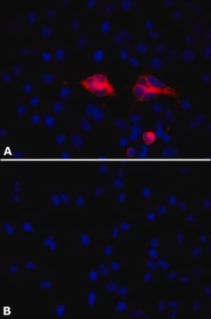
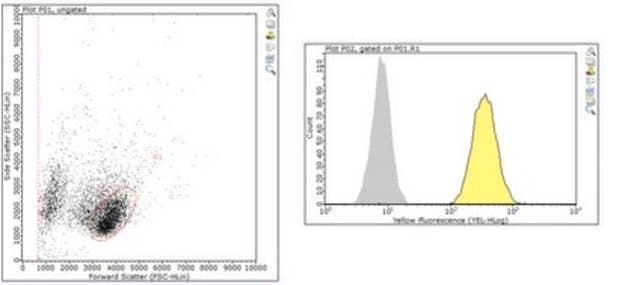
Flow Cytometry Analysis: A representative lot of this antibody detected uPAR in MDA-MB-231, H1299 and PC3 cells (see Harel, E., et al., 2014 UCSF Scientific Poster).
Flow Cytometry Analysis: A representative lot of this antibody detected uPAR in HEK293 cells stably expressing uPAR (Dureseti et al., (2010) JBC. 285(35):26878–26888).
Inhibition Assay: A representative lot of this antibody inhibted the invasion of MDA-MB-231 cells (see Harel, E., et al., 2014 UCSF Scientific Poster). Inhibition of uPAR is maximized with the combination of Anti-uPAR, clone 3C6 (Cat. # MABC89) and Anti-uPAR, clone 2G10 (Cat. # MABC88).
Inhibition Assay: A representative lot of this antibody blocked uPAR-mediated invasion and signaling in H1299 cells (Dureseti et al., (2010) JBC. 285(35):26878–26888). Inhibition of uPAR is maximized with the combination of Anti-uPAR, clone 3C6 (Cat. # MABC89) and Anti-uPAR, clone 2G10 (Cat. # MABC88).
品質
Evaluated by Western Blotting in reduced and non-reduced human uPAR recombinant protein.
Western Blotting Analysis: A 1:2500 dilution of this antibody detected uPAR in 1 µg of non-reduced human uPAR recombinant protein, but not in 1 µg of reduced human uPAR recombinant protein.
Western Blotting Analysis: A 1:2500 dilution of this antibody detected uPAR in 1 µg of non-reduced human uPAR recombinant protein, but not in 1 µg of reduced human uPAR recombinant protein.
ターゲットの説明
~45 kDa observed.
Recombinant human uPAR (Leu23-Arg303) has a predicted molecular weight of ~31 kDa, but runs around ~45 kDa under non-reduced conditions.
Recombinant human uPAR (Leu23-Arg303) has a predicted molecular weight of ~31 kDa, but runs around ~45 kDa under non-reduced conditions.
物理的形状
Protein A purified
Format: Purified
Purified rabbit monoclonal IgGλ in buffer containing PBS without preservatives.
保管および安定性
Stable for 1 year at -20°C from date of receipt.
Handling Recommendations: Upon receipt and prior to removing the cap, centrifuge the vial and gently mix the solution. Aliquot into microcentrifuge tubes and store at -20°C. Avoid repeated freeze/thaw cycles, which may damage IgG and affect product performance.
Handling Recommendations: Upon receipt and prior to removing the cap, centrifuge the vial and gently mix the solution. Aliquot into microcentrifuge tubes and store at -20°C. Avoid repeated freeze/thaw cycles, which may damage IgG and affect product performance.
その他情報
Concentration: Please refer to lot specific datasheet.
免責事項
Unless otherwise stated in our catalog or other company documentation accompanying the product(s), our products are intended for research use only and are not to be used for any other purpose, which includes but is not limited to, unauthorized commercial uses, in vitro diagnostic uses, ex vivo or in vivo therapeutic uses or any type of consumption or application to humans or animals.
適切な製品が見つかりませんか。
製品選択ツール.をお試しください
試験成績書(COA)
製品のロット番号・バッチ番号を入力して、試験成績書(COA) を検索できます。ロット番号・バッチ番号は、製品ラベルに「Lot」または「Batch」に続いて記載されています。
Moitza Principe et al.
Journal of hematology & oncology, 10(1), 16-16 (2017-01-15)
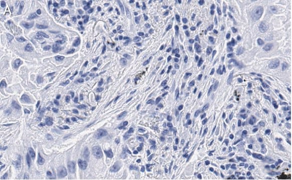
We have previously shown that in pancreatic ductal adenocarcinoma (PDA) cells, the glycolytic enzyme alpha-enolase (ENO1) also acts as a plasminogen receptor and promotes invasion and metastasis formation. Moreover, ENO1 silencing in PDA cells induces oxidative stress, senescence and profoundly
アクティブなフィルタ
ライフサイエンス、有機合成、材料科学、クロマトグラフィー、分析など、あらゆる分野の研究に経験のあるメンバーがおります。.
製品に関するお問い合わせはこちら(テクニカルサービス)