
M6319
Anti-Mitofusin-2 (N-Terminal) antibody produced in rabbit
affinity isolated antibody, buffered aqueous solution
Sinonimo/i:
Mitofusin 2 Antibody, Mitofusin 2 Antibody - Anti-Mitofusin-2 (N-Terminal) antibody produced in rabbit, Anti-CMT2A, Anti-CMT2A2, Anti-CPRP1, Anti-KIAA0214, Anti-MARF, Anti-Mfn2
Scegli un formato
| A voi/SKU | Disponibilità | Prezzo |
|---|---|---|
200 μL | Per informazioni sulla disponibilità, contatta il Servizio Clienti. | 566,00 € 481,10 € |
Informazioni su questo articolo
481,10 €
Prezzo di listino566,00 €Risparmia il 15%biological source
rabbit
Quality Segment
conjugate
unconjugated
antibody form
affinity isolated antibody
antibody product type
primary antibodies
clone
polyclonal
form
buffered aqueous solution
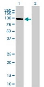
mol wt
antigen ~86 kDa
species reactivity
mouse, human, rat
technique(s)
immunoprecipitation (IP): 5-10 μg using HeLa human epitheloid carcinoma cell lysate, indirect immunofluorescence: 20-30 μg/mL using differentiated mouse C2 cells, western blot (chemiluminescent): 0.5-1 μg/mL using extracts of rat or mouse brain mitochondria
UniProt accession no.
shipped in
dry ice
storage temp.
−20°C
target post-translational modification
unmodified
Gene Information
human ... MFN2(9927)
mouse ... Mfn2(170731)
rat ... Mfn2(64476)
General description
Immunogen
Application
By immunoblotting, a working antibody concentration of 0.5-1 mg/mL is recommended using an extracts of rat and mouse brain mitochondria and a chemiluminescent detection reagent.
By indirect immunofluorescence, a working antibody concentration of 20-30 mg/mL is recommended using differentiated mouse C2 cells.
5-10 mg of the antibody immunoprecipitates mitofusin 2 from HeLa human epithelioid carcinoma cell lysate.
Western Blotting (1 paper)
Biochem/physiol Actions
Physical form
Disclaimer
1 of 1
Questo articolo | |||
|---|---|---|---|
| conjugate unconjugated | conjugate unconjugated | conjugate unconjugated | conjugate unconjugated |
| Quality Level 200 | Quality Level 100 | Quality Level 100 | Quality Level 100 |
| antibody form affinity isolated antibody | antibody form purified immunoglobulin | antibody form purified immunoglobulin | antibody form purified immunoglobulin |
| biological source rabbit | biological source mouse | biological source mouse | biological source mouse |
| Gene Information human ... MFN2(9927) | Gene Information human ... MFN2(9927) | Gene Information human ... MFN2(9927) | Gene Information human ... MFN1(55669) |
| species reactivity mouse, human, rat | species reactivity human, mouse, rat | species reactivity human, rat, mouse | species reactivity human |


Still not finding the right product?
Provate il nostro Motore di ricerca dei prodotti per restringere le opzioni.
Classe di stoccaggio
12 - Non Combustible Liquids
wgk
WGK 1
flash_point_f
Not applicable
flash_point_c
Not applicable
Scegli una delle versioni più recenti:
Possiedi già questo prodotto?
I documenti relativi ai prodotti acquistati recentemente sono disponibili nell’Archivio dei documenti.