
MABE1867
Anti-CLOCK Antibody, clone CLSP4
Sinonimo/i:
Circadian locomoter output cycles protein kaput, mCLOCK
About This Item
Prodotti consigliati


![CDP-Star® Disodium 2-chloro-5-(4-methoxyspiro {1,2-dioxetane-3,2′-(5′-chloro)tricyclo[3.3.1.13,7 ]decan}-4-yl)-1-phenyl phosphate](/deepweb/assets/sigmaaldrich/product/structures/224/846/d08963f5-c8ba-42e6-b4f7-9ee7fd76d809/640/d08963f5-c8ba-42e6-b4f7-9ee7fd76d809.png)


Origine biologica
mouse
Livello qualitativo
Forma dell’anticorpo
purified antibody
Tipo di anticorpo
primary antibodies
Clone
CLSP4, monoclonal
PM
calculated mol wt 96.39 kDa
observed mol wt ~N/A kDa
Purificato mediante
using protein G
Reattività contro le specie
mouse, human
Reattività contro le specie (prevista in base all’omologia)
rat
Confezionamento
antibody small pack of 100
tecniche
immunocytochemistry: suitable
immunofluorescence: suitable
immunohistochemistry: suitable
immunoprecipitation (IP): suitable
western blot: suitable
Isotipo
IgG1κ
Sequenza dell’epitopo
Internal
N° accesso ID proteina
N° accesso UniProt
Temperatura di conservazione
2-8°C
Informazioni sul gene
mouse ... Clock(12753)
Specificità
Immunogeno
Applicazioni
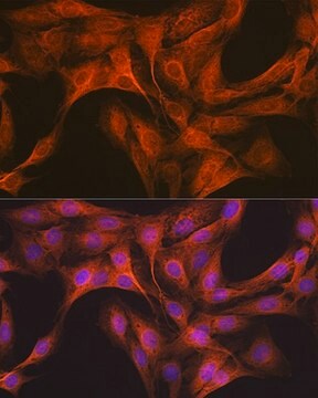
Evaluated by Immunocytochemistry in U2OS cells.
Immunocytochemistry Analysis: A 1:100 dilution of this antibody detected CLOCK in U2OS cells.
Tested Applications
Immunofluorescence Analysis: A representative lot detected CLOCK in Immunofluorescence applications (Umemura, Y., et al. (2017). Proc Natl Acad Sci USA. 114(36):E7479-E7488).
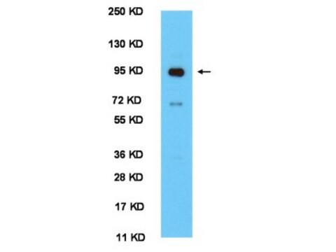
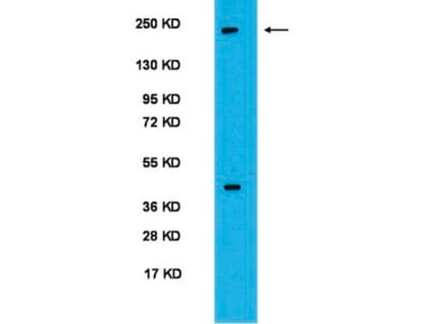
Western Blotting Analysis: A representative lot detected CLOCK in Western Blotting applications (Umemura, Y., et al. (2017). Proc Natl Acad Sci USA. 114(36):E7479-E7488).
Immunoprecipitation Analysis: A representative lot detected CLOCK in Immunoprecipitation applications (Yoshitane, H., et al. (2009). Mol Cell Biol. 29(13):3675-86).
Immunohistochemistry (Paraffin) Analysis: A representative lot detected CLOCK in Immunohistochemistry applications (Umemura, Y., et al. (2017). Proc Natl Acad Sci USA. 114(36):E7479-E7488).
Note: Actual optimal working dilutions must be determined by end user as specimens, and experimental conditions may vary with the end user.
Descrizione del bersaglio
Stato fisico
Ricostituzione
Stoccaggio e stabilità
Altre note
Esclusione di responsabilità
Non trovi il prodotto giusto?
Prova il nostro Motore di ricerca dei prodotti.
Codice della classe di stoccaggio
12 - Non Combustible Liquids
Classe di pericolosità dell'acqua (WGK)
WGK 1
Punto d’infiammabilità (°F)
Not applicable
Punto d’infiammabilità (°C)
Not applicable
Certificati d'analisi (COA)
Cerca il Certificati d'analisi (COA) digitando il numero di lotto/batch corrispondente. I numeri di lotto o di batch sono stampati sull'etichetta dei prodotti dopo la parola ‘Lotto’ o ‘Batch’.
Possiedi già questo prodotto?
I documenti relativi ai prodotti acquistati recentemente sono disponibili nell’Archivio dei documenti.
Il team dei nostri ricercatori vanta grande esperienza in tutte le aree della ricerca quali Life Science, scienza dei materiali, sintesi chimica, cromatografia, discipline analitiche, ecc..
Contatta l'Assistenza Tecnica.