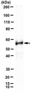
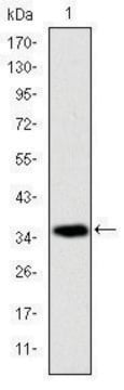
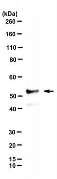
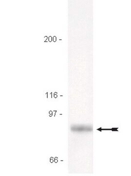
a peptide corresponding to amino acids 473-486 of murine RIP3.
Aplicación
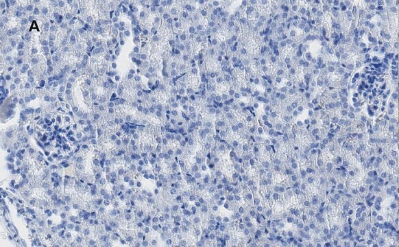
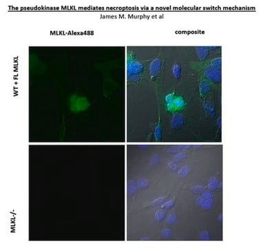
Applications in which this antibody has been used successfully, and the associated peer-reviewed papers, are given below. Immunocytochemistry (1 paper)
Ligadura / enlace
The action of this antibody can be blocked using blocking peptide SBP2283.
Forma física
Solution in phosphate buffered saline containing 0.02% sodium azide
Cláusula de descargo de responsabilidad
Unless otherwise stated in our catalog or other company documentation accompanying the product(s), our products are intended for research use only and are not to be used for any other purpose, which includes but is not limited to, unauthorized commercial uses, in vitro diagnostic uses, ex vivo or in vivo therapeutic uses or any type of consumption or application to humans or animals.
¿No encuentra el producto adecuado?
Pruebe nuestro Herramienta de selección de productos.
Exposure Limit Values (ELV) for artificial lighting were defined in order to prevent light-induced damage to the retina. The evaluation of the lighting devices include the correction of their spectra by the B(λ) function or blue light hazard function, representing
Acta biochimica et biophysica Sinica, 49(10), 879-889 (2017-10-06)
Receptor-interacting protein 3 (RIP3) is an essential component of the necroptosis signaling pathway. Phosphorylation of its downstream target, mixed lineage kinase domain-like protein (MLKL), has been proposed to induce necroptosis by initiating Ca2+ influx. Our previous studies have shown that
RIPK3 amyloid complex plays crucial roles during TNF-induced necroptosis and in response to immune defense in both human and mouse. Here, we have structurally characterized mouse RIPK3 homogeneous self-assembly using solid-state NMR, revealing a well-ordered N-shaped amyloid core structure featured
The receptor-interacting protein kinase 3 (RIPK3) is a key regulator of necroptosis and is involved in various pathologies of human diseases. We previously reported that RIPK3 expression is upregulated in various neural cells at the lesions and necroptosis contributed to
Duchenne muscular dystrophy (DMD) is a severe degenerative disorder caused by mutations in the dystrophin gene. Dystrophin-deficient muscles are characterised by progressive myofibre necrosis in which inflammation plays a deleterious role. However, the molecular mechanisms underlying inflammation-induced necrosis in muscle
Nuestro equipo de científicos tiene experiencia en todas las áreas de investigación: Ciencias de la vida, Ciencia de los materiales, Síntesis química, Cromatografía, Analítica y muchas otras.