Přejít k obsahu
Merck
Všechny fotografie(3)

Key Documents

L2164

Sigma-Aldrich

Anti-Luciferase antibody, Mouse monoclonal

enhanced validation

clone LUC-1, purified from hybridoma cell culture

Synonyma:

Anti-Luciferase Antibody

Přihlásitk zobrazení cen stanovených pro organizaci a smluvních cen


About This Item

MDL number:
UNSPSC Code:
12352203
NACRES:
NA.56

biological source

mouse

conjugate

unconjugated

antibody form

purified immunoglobulin

antibody product type

primary antibodies

clone

LUC-1, monoclonal

form

buffered aqueous solution

mol wt

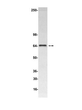
60 kDa

enhanced validation

recombinant expression
Learn more about Antibody Enhanced Validation

concentration

~2 mg/mL

technique(s)

immunocytochemistry: 20-40 μg/mL using transfected 293Tcells expressing luciferase
immunocytochemistry: suitable
western blot: 2-4 μg/mL using whole extract of transfected 293T cells expressing luciferase
western blot: suitable

shipped in

dry ice

storage temp.

−20°C

target post-translational modification

unmodified

Související kategorie

General description

Anti-Luciferase antibody, Mouse monoclonal , (mouse IgG1 isotype) is derived from the LUC-1 hybridoma produced by the fusion of mouse myeloma cells and splenocytes from BALB/c mice immunized with firefly (Photinus pyralis) luciferase. Luciferase, a reporter protein contains two binding pockets, one pocket for ATP and other one for luciferin substrate. Both the pockets are in close proximity to each other.
The mouse monoclonal Anti-Luciferase antibody recognizes the recombinant luciferase in transfected cells. This antibody provides user with an alternative detection method for luciferase. Since it directly detects the luciferase protein, it is not dependent on either the luciferase activity or the measeurement of rapid reaction kinetics. This also allows for the detection of the luciferase enzyme expression in situ with reproducibility between sample sets.

Specificity

Anti-Luciferase antibody, Mouse monoclonal recognizes recombinant luciferase in transfected eukaryotic (293T) cells.

Application

Anti-Luciferase antibody, Mouse monoclonal has been used in:
  • immunoblotting
  • immunocytochemistry
  • immunohistochemistry

Biochem/physiol Actions

Luciferase catalyzes a bioluminescent reaction in the presence of substrate luciferin as well as Mg2+, oxygen and ATP. The luciferase assay is rapid, sensitive, and unlike the CAT assay, does not require a radioactive substrate. Anti-Luciferase antibody can detect the luciferase enzyme expression in situ, providing a means to study the cellular localization of the signal sequence activation.

Physical form

Solution in 0.01 M phosphate buffered saline, pH 7.4, containing 15 mM sodium azide.

Preparation Note

Working Dilution: 1-2 μg/ml

Storage and Stability

For continuous use store at 2-8 °C for up to one month. For extended storage, freeze in working aliquots. Repeated freezing and thawing, or storage in a frost-free freezer, is not recommended. If slight turbidity occurs upon prolonged storage, clarify the solution by centrifugation before use. Working dilutions should be discarded if not used within 12 hours.

Disclaimer

Unless otherwise stated in our catalog, our products are intended for research use only and are not to be used for any other purpose, which includes but is not limited to, unauthorized commercial uses, in vitro diagnostic uses, ex vivo or in vivo therapeutic uses or any type of consumption or application to humans or animals.

Ještě jste nenalezli správný produkt?  

Vyzkoušejte náš produkt Nástroj pro výběr produktů.

Storage Class

10 - Combustible liquids

wgk_germany

WGK 1

flash_point_f

Not applicable

flash_point_c

Not applicable


Osvědčení o analýze (COA)

Vyhledejte osvědčení Osvědčení o analýze (COA) zadáním čísla šarže/dávky těchto produktů. Čísla šarže a dávky lze nalézt na štítku produktu za slovy „Lot“ nebo „Batch“.

Již tento produkt vlastníte?

Dokumenty související s produkty, které jste v minulosti zakoupili, byly za účelem usnadnění shromážděny ve vaší Knihovně dokumentů.

Navštívit knihovnu dokumentů

Sung-Yong Kim et al.
Plant physiology, 142(3), 984-992 (2006-09-12)
Extracellular ATP (eATP) in animals is well documented and known to play an important role in cellular signaling (e.g. at the nerve synapse). The existence of eATP has been postulated in plants; however, there is no definitive experimental evidence for
Dun Jack Fu et al.
Translational vision science & technology, 9(13), 44-44 (2021-01-15)
The purpose of this study was to develop and characterize a novel bioluminescence transgenic mouse model that facilitates rapid evaluation of genetic medicine delivery methods for inherited and acquired corneal diseases. Corneal expression of the firefly luciferase transgene (luc2) was
Jie Gao et al.
Plant signaling & behavior, 16(7), 1916211-1916211 (2021-05-27)
Aluminum (Al) toxicity in acidic soils severely reduces crop production worldwide. Sorghum (Sorghum bicolor L.) is an important agricultural crop widely grown in tropical and subtropical regions, where Al toxicity is prevalent. ATP-binding cassette (ABC) transporters play key roles in
Megan Culler Freeman et al.
Journal of virology, 88(10), 5319-5327 (2014-03-14)
The replication of coronaviruses occurs in association with multiple virus-induced membrane structures that evolve during the course of infection; however, the dynamics of this process remain poorly understood. Previous studies of coronavirus replication complex organization and protein interactions have utilized
In vivo natriuretic peptide reporter assay identifies chemical modifiers of hypertrophic cardiomyopathy signalling
Becker JR, et al.
Cardiovascular Research, 93(3), 463-470 (2012)

Náš tým vědeckých pracovníků má zkušenosti ve všech oblastech výzkumu, včetně přírodních věd, materiálových věd, chemické syntézy, chromatografie, analytiky a mnoha dalších..

Obraťte se na technický servis.