Přejít k obsahu
Merck
Všechny fotografie(4)

Key Documents

C1821

Sigma-Aldrich

Monoclonal Anti-Pan Cadherin antibody produced in mouse

clone CH-19, ascites fluid

Synonyma:

Monoclonal Anti-Pan Cadherin

Přihlásitk zobrazení cen stanovených pro organizaci a smluvních cen


About This Item

UNSPSC Code:
12352203
NACRES:
NA.41

biological source

mouse

conjugate

unconjugated

antibody form

ascites fluid

antibody product type

primary antibodies

clone

CH-19, monoclonal

mol wt

antigen 135 kDa

contains

15 mM sodium azide

species reactivity

canine, frog, snake, guinea pig, chicken, pig, feline, bovine, rabbit, hamster, Psammomys (sand rat), goat, rat, human, sheep, mouse

technique(s)

immunohistochemistry (formalin-fixed, paraffin-embedded sections): 1:500 using protease-digested animal heart sections
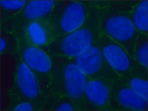
indirect immunofluorescence: 1:500 using cultured MDBK cells
microarray: suitable
western blot: suitable

isotype

IgG1

UniProt accession no.

application(s)

research pathology

shipped in

dry ice

storage temp.

−20°C

target post-translational modification

unmodified

Hledáte podobné produkty? Navštivte Průvodce porovnáváním produktů

General description

Cadherins are members of a multigene family of single chain glycoprotein receptors mediating Ca2+ -dependent cell-cell adhesion. The N-terminal part of these molecule is exposed on the external cell surface and contains the putative homophilic binding sites. This is followed by a typical single transmembrane sequence and usually a cytoplasmic C-terminal tail which mediates interaction with the microfilament system through molecules such as catenins, plakoglobin, vinculin, and α-actinin.
Cadherins that are primarily located in areas of cell-cell contacts are involved in selective cell sorting and in the mechanical cytoplasmic response. They are implicated in morphogenetic processes, intercellular signalling and in tumor invasiveness and metastasis.
Multiple cadherins have been characterized from diverse species and tissues including E-Cadherin, N-Cadherin (A-CAM), P-Cadherin, V-Cadherin, R-Cadherin and T-Cadherin. Specific polyclonal antibodies against a highly conserved sequence from the cytoplasmic C-terminal of N-Cadherin has been prepared.
Neural-cadherin (N-cadherin) is a member of the classic cadherin family. It is mainly expressed in neural and mesenchymal tissues.

Specificity

Detection of members of the cadherin family or genetically engineered proteins containing the C-terminal cadherin tail, stains adherens type cell-cell junctions regardless of their cadherin type.
Monoclonal Anti-Pan Cadherin antibody reacts extensively with all known members of the cadherin family. The cadherins migrate in the molecular weight range of 125 to 140 kDa and are tissue dependent. The antibody reacts using frozen sections, protease-digested, formalin-fixed paraffin-embedded, Bouin′s-fixed and acetone-fixed tissue sections. It reacts with cultured cell line preparations. Cross-reaction has been observed with human, rabbit, bovine, goat, sheep, dog, pig, guinea pig, hamster, cat, rat, mouse, psammomys, chicken, frog and snake.

Immunogen

synthetic peptide corresponding to the C-terminal amino acids of chicken N-Cadherin with an extra N-terminal lysine residue (24 amino acids) coupled to KLH.

Application

Monoclonal Anti-Pan Cadherin antibody produced in mouse has been used in:
  • fluorescence imaging
  • immunohistochemistry
  • western blotting
  • immunoblotting
  • immunofluorescence

Monoclonal Anti-Pan Cadherin may be used for: immunochemical and immunocytochemical detection of members of the cadherin family in normal and neoplastic cells and tissues; identification of genetically engineered proteins containing the C-terminal cadherin tail; screening of cDNA expression libraries for identification of novel members of the cadherin family and demonstration of adherens-type cell-cell junctions irrespective of their cadherin subtype.

Biochem/physiol Actions

Neural-cadherin (N-cadherin) has an oncogenic role in bladder cancer, breast cancer, prostate cancer and melanoma. It stimulates the heterotypic adhesion of melanoma cells to dermal fibroblast cells. N-cadherin acts as a critical modulator for the migration of neuronal progenitors. It is essential for the proper cohesion and polarisation of neuroepithelial cells. N-cadherin is important for normal gastrulation and neural crest development. It is required for myogenesis and myotube formation.

Disclaimer

Unless otherwise stated in our catalog or other company documentation accompanying the product(s), our products are intended for research use only and are not to be used for any other purpose, which includes but is not limited to, unauthorized commercial uses, in vitro diagnostic uses, ex vivo or in vivo therapeutic uses or any type of consumption or application to humans or animals.

Ještě jste nenalezli správný produkt?  

Vyzkoušejte náš produkt Nástroj pro výběr produktů.

recommended

Storage Class

12 - Non Combustible Liquids

wgk_germany

nwg

flash_point_f

Not applicable

flash_point_c

Not applicable


Osvědčení o analýze (COA)

Vyhledejte osvědčení Osvědčení o analýze (COA) zadáním čísla šarže/dávky těchto produktů. Čísla šarže a dávky lze nalézt na štítku produktu za slovy „Lot“ nebo „Batch“.

Již tento produkt vlastníte?

Dokumenty související s produkty, které jste v minulosti zakoupili, byly za účelem usnadnění shromážděny ve vaší Knihovně dokumentů.

Navštívit knihovnu dokumentů

N-cadherin and fibroblast growth factor receptors crosstalk in the control of developmental and cancer cell migrations
Nguyen T and Mege RM
European Journal of Cell Biology, 95(11), 415-426 (2016)
N-cadherin promotes thyroid tumorigenesis through modulating major signaling pathways
Da C, et al.
Oncotarget, 8(5), 8131-8131 (2017)
Expression of cadherin adhesion molecules on human gametes
Rufas O, et al.
Molecular Human Reproduction, 6(2), 163-169 (2000)
GRP78 and Cripto form a complex at the cell surface and collaborate to inhibit transforming growth factor beta signaling and enhance cell growth
Shani G, et al.
Molecular and Cellular Biology, 28(2), 666-677 (2008)
Sung Jun Bae et al.
eLife, 6 (2017-10-25)
The Hippo pathway controls tissue growth and homeostasis through a central MST-LATS kinase cascade. The scaffold protein SAV1 promotes the activation of this kinase cascade, but the molecular mechanisms remain unknown. Here, we discover SAV1-mediated inhibition of the PP2A complex

Náš tým vědeckých pracovníků má zkušenosti ve všech oblastech výzkumu, včetně přírodních věd, materiálových věd, chemické syntézy, chromatografie, analytiky a mnoha dalších..

Obraťte se na technický servis.