MAB3420
Anti-Tau-1 Antibody, clone PC1C6
clone PC1C6, Chemicon®, from mouse
Synonyma:
Anti-Tau Antibody
About This Item
Doporučené produkty
biological source
mouse
Quality Level
antibody form
purified antibody
clone
PC1C6, monoclonal
species reactivity
human, rat, bovine
packaging
antibody small pack of 25 μg
manufacturer/tradename
Chemicon®
technique(s)
immunofluorescence: suitable
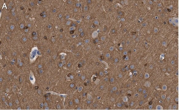
immunohistochemistry: suitable
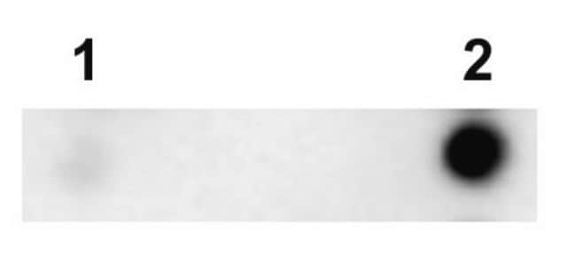
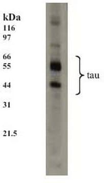
western blot: suitable
isotype
IgG2a
NCBI accession no.
UniProt accession no.
shipped in
dry ice
storage temp.
−20°C
target post-translational modification
unmodified
Související kategorie
General description
Specificity
Immunogen
Application
Neuroscience
Neurodegenerative Diseases
Immunofluorescence: A 1:1000 dilution of this antibody detected Tau in mouse primary neurons. (Basnet, N., et al. (2018). Nat. Cell Biol. 20(10); 1172-1180.
Immunohistochemistry: 5 μg/mL; stains axons in tissue primarily, however in culture Tau expression is not restricted to just axons.
Optimal working dilutions must be determined by end user.
Immunohistochemistry Protocol
Dephosphorylation of tissue sections (optional)
Dephosphorylation with alkaline phosphatase is recommended for staining neurofibrillary tangles in Alzheimer′s brain tissue with anti-tau-1 (6). This treatment changes the staining pattern of anti-tau-1 to include cell bodies, dendrites and axons of neurons. In untreated samples, anti-tau-1 stains axons only.
1. Incubate tissue sections at +32°C for 2.5 hours with constant agitation in the following solution: 100 mM Tris-HCl, pH 8.0; 130 units/mL alkaline phosphatase, 1 mM PMSF, 10 μg/mL pepstatin and 10 μg/mL leupeptin.
2. Rinse sections twice, 3 min per rinse, with 100 mM Tris-HCl, pH 8.0.
Anti-tau-1 staining
1. Block non-specific binding by incubating sections in PBS containing 1% (v/v) normal animal serum, and 0.03% (w/v) Triton X-100. The animal serum should be from the same species as the secondary antibody.
2. Rinse 3 times with PBS, 3 min per rinse.
3. Incubate sections with anti-tau-1, approximately 5 μg/mL, diluted in PBS containing 1% (v/v) normal animal serum.
4. Wash with PBS, changing the solution 3 times over a 3 min period.
5. Detect with a standard secondary antibody detection system (10-13).
Target description
Linkage
Physical form
Storage and Stability
Analysis Note
Alzheimer′s brain tissue (dephosphorylation with alkaline phosphatase is recommended for staining neurofibrillary tangles in Alzheimer’s brain tissue) or human T98G glioblastoma cells
Other Notes
Legal Information
Disclaimer
recommended
Storage Class
12 - Non Combustible Liquids
wgk_germany
WGK 2
flash_point_f
Not applicable
flash_point_c
Not applicable
Osvědčení o analýze (COA)
Vyhledejte osvědčení Osvědčení o analýze (COA) zadáním čísla šarže/dávky těchto produktů. Čísla šarže a dávky lze nalézt na štítku produktu za slovy „Lot“ nebo „Batch“.
Již tento produkt vlastníte?
Dokumenty související s produkty, které jste v minulosti zakoupili, byly za účelem usnadnění shromážděny ve vaší Knihovně dokumentů.
Zákazníci si také prohlíželi
Sortimentní položky
Immunofluorescence uses antibody-conjugated fluorescent molecules for protein localization, modification confirmation, and protein complex visualization.
Derivation and characterization of functional human neural stem cell derived oligodendrocyte progenitor cells (OPCs) that efficiently myelinate primary neurons in culture.
Protokoly
Tips and troubleshooting for FFPE and frozen tissue immunohistochemistry (IHC) protocols using both brightfield analysis of chromogenic detection and fluorescent microscopy.
Náš tým vědeckých pracovníků má zkušenosti ve všech oblastech výzkumu, včetně přírodních věd, materiálových věd, chemické syntézy, chromatografie, analytiky a mnoha dalších..
Obraťte se na technický servis.


