Přejít k obsahu
Merck
Všechny fotografie(1)

Key Documents

FCMAB168P

Sigma-Aldrich

Milli-Mark Anti-CD3 -PE Antibody, clone UCHT1

clone UCHT1, Milli-Mark®, from mouse

Synonyma:

T-cell receptor

Přihlásitk zobrazení cen stanovených pro organizaci a smluvních cen


About This Item

UNSPSC Code:
12352203
eCl@ss:
32160702
NACRES:
NA.44

biological source

mouse

Quality Level

conjugate

PE

antibody form

purified immunoglobulin

antibody product type

primary antibodies

clone

UCHT1, monoclonal

species reactivity

human

manufacturer/tradename

Milli-Mark®

technique(s)

flow cytometry: suitable

isotype

IgG1κ

NCBI accession no.

UniProt accession no.

shipped in

wet ice

target post-translational modification

unmodified

Gene Information

human ... CD3E(916)

General description

Human TCR/CD3 is a complex structure on the lymphocyte surface. It consists of the TCR alpha beta or TCR gamma delta heterodimer and the associated CD3 complex. The CD3 complex is composed of six polypeptides with usually four different transmembrane CD3 chains, gamma, delta, epsilon. and zeta. Three different dimers, gamma epsilon, delta epsilon, and zeta zeta, constitute the CD3 complex. The MW of CD3epsilon is 20 000 (Campana, 1987). CD3 is first detectable in early thymocytes and its appearance probably represents one of the earliest signs of commitment to the T cell lineage. In cortical thymocytes, during early stages of maturation, the CD3 antigen is predominantly present in the cell cytoplasm. In medullary thymocyles, the CD3 antigen is predominantly detected on the cell surface (Swerdlow, 1988; Beverley, 1981). The majority of T cell neoplasms also expresses CD3, but it is absent from non-T-cell lymphoid malignancies (McMichael, 1987). Consistent with the pattern of synthesis of the antigen in normal thymocytes, the earliest site detectable within neoplastic cells is the cell cytoplasm (Swerdlow, 1988).

Specificity

Antibody recognizes Human CD3 antigen.

Immunogen

Human infant thymocytes and lymphocytes from a patient with Sézary disease.

Application

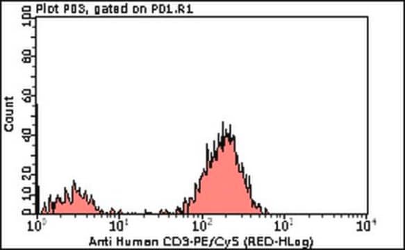
Milli-Mark Anti-CD3 -PE Antibody, clone UCHT1 is an antibody against CD3 for use in FC.
Research Category
Inflammation & Immunology
Research Sub Category
Immunoglobulins & Immunology

Quality

Evaluated by flow cytometry using Human Lymphocytes.

Target description

20 kDa (Campana, 1987)

Physical form

Protein A purified
Purified Mouse monoclonal IgG1 conjugated to PE in 0.05M Tris-HCl buffer with 1% BSA, Sodium Azide at 0.097% at pH 7.2

Storage and Stability

Maintain refrigerated at 2-8 °C protected from light in undiluted aliquots for up to 6 months from date of receipt.

Analysis Note

Control
Human Lymphocytes

Other Notes

Concentration: Please refer to the Certificate of Analysis for the lot-specific concentration.

Legal Information

MILLI-MARK is a registered trademark of Merck KGaA, Darmstadt, Germany

Disclaimer

Unless otherwise stated in our catalog or other company documentation accompanying the product(s), our products are intended for research use only and are not to be used for any other purpose, which includes but is not limited to, unauthorized commercial uses, in vitro diagnostic uses, ex vivo or in vivo therapeutic uses or any type of consumption or application to humans or animals.

Ještě jste nenalezli správný produkt?  

Vyzkoušejte náš produkt Nástroj pro výběr produktů.

Storage Class

10 - Combustible liquids

wgk_germany

WGK 2

flash_point_f

Not applicable

flash_point_c

Not applicable


Osvědčení o analýze (COA)

Vyhledejte osvědčení Osvědčení o analýze (COA) zadáním čísla šarže/dávky těchto produktů. Čísla šarže a dávky lze nalézt na štítku produktu za slovy „Lot“ nebo „Batch“.

Již tento produkt vlastníte?

Dokumenty související s produkty, které jste v minulosti zakoupili, byly za účelem usnadnění shromážděny ve vaší Knihovně dokumentů.

Navštívit knihovnu dokumentů

Náš tým vědeckých pracovníků má zkušenosti ve všech oblastech výzkumu, včetně přírodních věd, materiálových věd, chemické syntézy, chromatografie, analytiky a mnoha dalších..

Obraťte se na technický servis.