Saltar al contenido
Merck
Todas las fotos(1)

Key Documents

ABT170

Sigma-Aldrich

Anti-alpha Tubulin Antibody, nontyrosinated

serum, from rabbit

Sinónimos:

Tubulin alpha-1A chain, Alpha-tubulin 3, Tubulin B-alpha-1, Tubulin alpha-3 chain

Iniciar sesiónpara Ver la Fijación de precios por contrato y de la organización


About This Item

UNSPSC Code:
12352203
eCl@ss:
32160702
NACRES:
NA.41

biological source

rabbit

Quality Level

antibody form

serum

antibody product type

primary antibodies

clone

polyclonal

species reactivity

mouse, porcine

species reactivity (predicted by homology)

rat (based on 100% sequence homology), human (based on 100% sequence homology)

technique(s)

ELISA: suitable
immunocytochemistry: suitable
western blot: suitable

NCBI accession no.

UniProt accession no.

shipped in

wet ice

target post-translational modification

unmodified

Gene Information

human ... TUBA1A(7846)

General description

Microtubules are involved in a wide variety of cellular activities ranging from mitosis and transport events to cell movement and the maintenance of cell shape. Tubulin itself is a globular protein which consists of two polypeptides (alpha and beta tubulin). Alpha and beta tubulin dimers are assembled to 13 protofilaments that form a microtubule of 22 nm diameter. Tyrosine ligase adds a C-terminal tyrosin to monomeric alpha tubulin. Assembled microtubules can be detyrosinated by a cytoskeleton associated carboxypeptidase. Detyrosinated alpha tubulin is referred to as Glu-tubulin. Another post-translational modification of detyrosinated alpha tubulin is C-terminal polyglutamylation which is characteristic for microtubules in neuronal cells and the mitotic spindle.

Specificity

This antibody detects the nontyrosinated form of alpha Tubulin.

Immunogen

KLH-conjugated linear peptide corresponding to human nontyrosinated alpha Tubulin.

Application

Detect Tubulin using this rabbit polyclonal antibody, Anti-alpha Tubulin Antibody, nontyrosinated validated for use in western blotting, ELISA & ICC.
Research Category
Cell Structure
Research Sub Category
Cytoskeleton
Western Blotting Analysis: A representative lot from an independent laboratory specifically detected nontyrosinated alpha Tubulin and not tyrosinated alpha Tubulin from purified porcine alpha Tubulin (Gundersen, G. G., et al. (1984). Cell. 38(3):779-89.).

ELISA Analysis: A representative lot from an independent laboratory specifically detected nontyrosinated alpha Tubulin, and not nontyrosinated alpha Tubulin in an indirect ELISA and in a competitive ELISA (Gundersen, G. G., et al. (1984). Cell. 38(3):779-89.).

Immunocytochemistry Analysis: A representative lot from an independent laboratory specifically detected nontyrosinated alpha Tubulin, and not nontyrosinated alpha Tubulin in TC-7 cells in interphase (Gundersen, G. G., et al. (1984). Cell. 38(3):779-89.).

Quality

Evaluated by Western Blotting in NIH/3T3 cell lysate, which were untreated or treated with Pancreatic Carboxypeptidase (PCA).

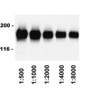
Western Blotting Analysis: A 1:2,000 dilution of this antibody detected alpha Tubulin, nontyrosinated in 10 µg of PCA treated NIH/3T3 cell lysate and demonstrated a loss of signal in untreated NIH/3T3 cell lysate.

Target description

~51 kDa observed

Linkage

Replaces: 04-1117

Physical form

Rabbit polyclonal serum in buffer containing serum with 0.05% sodium azide.
Unpurified

Storage and Stability

Stable for 1 year at -20°C from date of receipt.
Handling Recommendations: Upon receipt and prior to removing the cap, centrifuge the vial and gently mix the solution. Aliquot into microcentrifuge tubes and store at -20°C. Avoid repeated freeze/thaw cycles, which may damage IgG and affect product performance.

Disclaimer

Unless otherwise stated in our catalog or other company documentation accompanying the product(s), our products are intended for research use only and are not to be used for any other purpose, which includes but is not limited to, unauthorized commercial uses, in vitro diagnostic uses, ex vivo or in vivo therapeutic uses or any type of consumption or application to humans or animals.

¿No encuentra el producto adecuado?  

Pruebe nuestro Herramienta de selección de productos.

Storage Class

10 - Combustible liquids

wgk_germany

WGK 1


Certificados de análisis (COA)

Busque Certificados de análisis (COA) introduciendo el número de lote del producto. Los números de lote se encuentran en la etiqueta del producto después de las palabras «Lot» o «Batch»

¿Ya tiene este producto?

Encuentre la documentación para los productos que ha comprado recientemente en la Biblioteca de documentos.

Visite la Librería de documentos

Hui Li et al.
Frontiers in psychiatry, 9, 588-588 (2018-11-30)
Estrogen (E2) withdrawal is a core pathology mechanism for postpartum depression (PPD). Galanin (GAL), an estrogen-inducible neuropeptide has also been reported to be associated with depression. However, it still remains unclear which GAL receptors (GALRs) are involved in PPD pathologic
Lei Feng et al.
Developmental biology, 395(2), 268-286 (2014-09-23)
L-fucose, a monosaccharide widely distributed in eukaryotes and certain bacteria, is a determinant of many functional glycans that play central roles in numerous biological processes. The molecular mechanism, however, by which fucosylation mediates these processes remains largely elusive. To study
Saifeng Cheng et al.
Cell, 185(17), 3169-3185 (2022-08-01)
Mice deficient for all ten-eleven translocation (TET) genes exhibit early gastrulation lethality. However, separating cause and effect in such embryonic failure is challenging. To isolate cell-autonomous effects of TET loss, we used temporal single-cell atlases from embryos with partial or
Marko Dunjić et al.
Nature communications, 14(1), 3791-3791 (2023-06-27)
Eviction of histones from nucleosomes and their exchange with newly synthesized or alternative variants is a central epigenetic determinant. Here, we define the genome-wide occupancy and exchange pattern of canonical and non-canonical histone variants in mouse embryonic stem cells by
Histone deacetylase inhibitors (HDACi) increase expression of KCa2.3 (SK3) in primary microvascular endothelial cells.
Kolski-Andreaco, et al.
American Journal of Physiology. Cell Physiology, 322, C338-C353 (2023)

Nuestro equipo de científicos tiene experiencia en todas las áreas de investigación: Ciencias de la vida, Ciencia de los materiales, Síntesis química, Cromatografía, Analítica y muchas otras.

Póngase en contacto con el Servicio técnico