
T5530
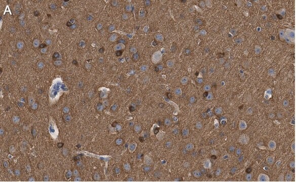
ANTI-TAU (MAPT) Antibody
mouse monoclonal, TAU-2
Sélectionner une taille de conditionnement
606.00 CHF
Date d'expédition estimée le01 mai 2025
Sélectionner une taille de conditionnement
About This Item
606.00 CHF
Date d'expédition estimée le01 mai 2025
Produits recommandés


Nom du produit
Monoclonal Anti-τ (Tau) antibody produced in mouse, clone TAU-2, ascites fluid
Source biologique
mouse
Niveau de qualité
Conjugué
unconjugated
Forme d'anticorps
ascites fluid
Type de produit anticorps
primary antibodies
Clone
TAU-2, monoclonal
Poids mol.
antigen 55-62 kDa
Contient
15 mM sodium azide
Espèces réactives
monkey, bovine, chicken, human
Technique(s)
immunohistochemistry (formalin-fixed, paraffin-embedded sections): suitable
microarray: suitable
western blot: 1:1,000 using a fresh total bovine brain extract or an enriched microtubule protein preparation
Isotype
IgG1
Numéro d'accès UniProt
Conditions d'expédition
dry ice
Température de stockage
−20°C
Modification post-traductionnelle de la cible
unmodified
Informations sur le gène
human ... MAPT(4137)
Catégories apparentées
Description générale
Immunogène
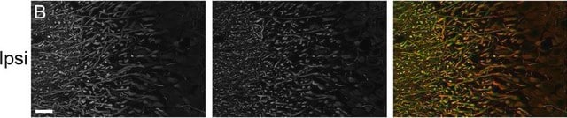
Application
- in immunohistology
- in immunoblotting
- in dot blot
- in immunohistochemistry
Actions biochimiques/physiologiques
Clause de non-responsabilité
Vous ne trouvez pas le bon produit ?
Essayez notre Outil de sélection de produits.
Code de la classe de stockage
10 - Combustible liquids
Classe de danger pour l'eau (WGK)
WGK 3
Point d'éclair (°F)
Not applicable
Point d'éclair (°C)
Not applicable
Faites votre choix parmi les versions les plus récentes :
Certificats d'analyse (COA)
Vous ne trouvez pas la bonne version ?
Si vous avez besoin d'une version particulière, vous pouvez rechercher un certificat spécifique par le numéro de lot.
Déjà en possession de ce produit ?
Retrouvez la documentation relative aux produits que vous avez récemment achetés dans la Bibliothèque de documents.
Active Filters
Notre équipe de scientifiques dispose d'une expérience dans tous les secteurs de la recherche, notamment en sciences de la vie, science des matériaux, synthèse chimique, chromatographie, analyse et dans de nombreux autres domaines..
Contacter notre Service technique