Direkt zum Inhalt
Merck

07-502-I

Sigma-Aldrich

Anti-p67-phox (not Human) Antibody

from rabbit, purified by affinity chromatography

Synonym(e):

Neutrophil cytosol factor 2, NCF-2, 67 kDa neutrophil oxidase factor, NADPH oxidase activator 2, Neutrophil NADPH oxidase factor 2, p67-phox, p67-phox (rodent)

Anmeldenzur Ansicht organisationsspezifischer und vertraglich vereinbarter Preise


About This Item

UNSPSC-Code:
12352203
eCl@ss:
32160702
NACRES:
NA.41

Biologische Quelle

rabbit

Qualitätsniveau

Antikörperform

affinity isolated antibody

Antikörper-Produkttyp

primary antibodies

Klon

polyclonal

Aufgereinigt durch

affinity chromatography

Speziesreaktivität

rat, mouse

Methode(n)

immunohistochemistry: suitable
western blot: suitable

NCBI-Hinterlegungsnummer

UniProt-Hinterlegungsnummer

Versandbedingung

wet ice

Posttranslationale Modifikation Target

unmodified

Angaben zum Gen

mouse ... Ncf2(17970)
rat ... Ncf2(364018)

Allgemeine Beschreibung

Neutrophil cytosol factor 2 (UniProt Q09014; also known as 67 kDa neutrophil oxidase factor, NADPH oxidase activator 2, NADPH oxidase subunit 67 kDa, NCF-2, Ncf2, Neutrophil NADPH oxidase factor 2, p67-phox, p47phox) is encoded by the murine Ncf2 (also known as Noxa2, p67phox) gene in murine species (Gene ID 17970). Activation of NADPH-oxidase dependent superoxide generation requires the participation of several cytosolic factors, including p47-phox, p67-phox, p40-phox and Rac. In the resting cell p47phox, p67phox, and p40phox form a large molecular weight cytosolic complex. Activation of the NADPH oxidase is initiated by the assembly of p47phox, p67phox, and Rac with cytochrome b558 in a 1:1:1:1 complex in the plasma membrane.

Immunogen

Epitope: N-terminus
KLH-conjugated linear peptide corresponding to the N-terminus of mouse p67-phox.

Anwendung

Research Category
Neurowissenschaft
Research Sub Category
Entwicklungsabhängige Signalübertragung
Anti-p67-phox (not Human) Antibody is an antibody against p67-phox, tested in mouse and rat, but is not expected to react with human, for use in Western Blotting and Immunohistochemistry.
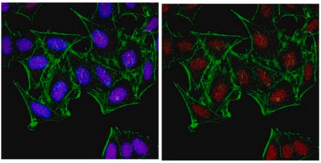
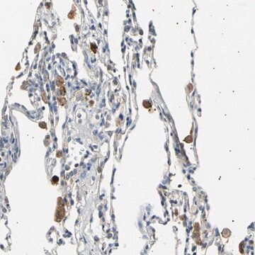
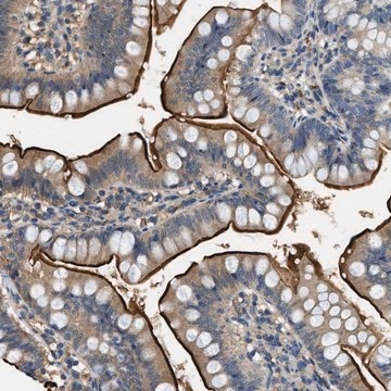
Immunohistochemistry Analysis: A 1:50 dilution from a representative lot detected p67-phox (rodent) in rat colon and mouse hindbrain tissue.

Qualität

Evaluated by Western Blotting in C6 and THP-1 cell lysate.

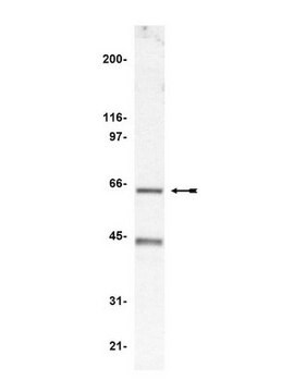
Western Blotting Analysis: 0.5 µg/mL of this antibody detected p67-phox (rodent) in 10 µg of C6 and THP-1 cell lysate.

Zielbeschreibung

~72 kDa observed. Uncharacterized band(s) may appear in some lysates.

Physikalische Form

Affinity purified
Purified rabbit polyclonal antibody in buffer containing 0.1 M Tris-Glycine (pH 7.4), 150 mM NaCl with 0.05% sodium azide.

Lagerung und Haltbarkeit

Stable for 1 year at 2-8°C from date of receipt.

Sonstige Hinweise

Concentration: Please refer to lot specific datasheet.

Haftungsausschluss

Unless otherwise stated in our catalog or other company documentation accompanying the product(s), our products are intended for research use only and are not to be used for any other purpose, which includes but is not limited to, unauthorized commercial uses, in vitro diagnostic uses, ex vivo or in vivo therapeutic uses or any type of consumption or application to humans or animals.

Not finding the right product?  

Try our Produkt-Auswahlhilfe.

Lagerklassenschlüssel

12 - Non Combustible Liquids

WGK

WGK 1

Flammpunkt (°F)

Not applicable

Flammpunkt (°C)

Not applicable


Analysenzertifikate (COA)

Suchen Sie nach Analysenzertifikate (COA), indem Sie die Lot-/Chargennummer des Produkts eingeben. Lot- und Chargennummern sind auf dem Produktetikett hinter den Wörtern ‘Lot’ oder ‘Batch’ (Lot oder Charge) zu finden.

Besitzen Sie dieses Produkt bereits?

In der Dokumentenbibliothek finden Sie die Dokumentation zu den Produkten, die Sie kürzlich erworben haben.

Die Dokumentenbibliothek aufrufen

Unser Team von Wissenschaftlern verfügt über Erfahrung in allen Forschungsbereichen einschließlich Life Science, Materialwissenschaften, chemischer Synthese, Chromatographie, Analytik und vielen mehr..

Setzen Sie sich mit dem technischen Dienst in Verbindung.