Accéder au contenu
Merck
Toutes les photos(2)

Key Documents

H0412

Sigma-Aldrich

Anti-phospho-Histone H3 (pSer10) antibody produced in rabbit

IgG fraction of antiserum, buffered aqueous solution

Synonyme(s) :

PH3 Antibody - Anti-phospho-Histone H3 (pSer10) antibody produced in rabbit, Ph3 Antibody, Anti-H3S10p

Se connecterpour consulter vos tarifs contractuels et ceux de votre entreprise/organisme


About This Item

Numéro MDL:
Code UNSPSC :
12352203
Nomenclature NACRES :
NA.41

Source biologique

rabbit

Conjugué

unconjugated

Forme d'anticorps

IgG fraction of antiserum

Type de produit anticorps

primary antibodies

Clone

polyclonal

Forme

buffered aqueous solution

Poids mol.

antigen 17 kDa

Espèces réactives

Caenorhabditis elegans, bovine, human, rat, chicken, Drosophila, frog, mouse

Technique(s)

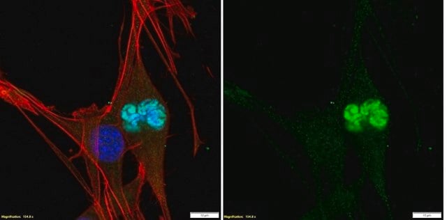
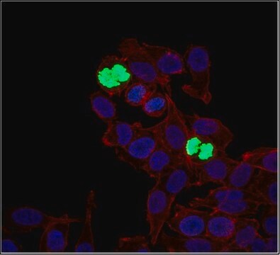
indirect immunofluorescence: 1:500 using human epitheloid carcinoma HeLa cell line treated with nocodazole.
microarray: suitable
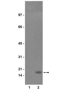
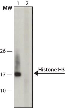
western blot: 1:2,000 using whole cell extract of human acute T cell leukemia Jurkat cell line treated with nocodazole.

Numéro d'accès UniProt

Conditions d'expédition

dry ice

Température de stockage

−20°C

Modification post-traductionnelle de la cible

phosphorylation (pSer10)

Description générale

Monoclonal Anti-Phosphoserine (mouse IgG1 isotype) is derived from the PSR-45 hybridoma produced by the fusion of mouse myeloma cells and splenocytes from a BALB/c mouse immunized with phosphoserine conjugated to KLH. Histone proteins H3, H4, H2A, H2B and H1 function as building blocks to package eukaryotic DNA into repeating nucleosome units that are folded in higher order chromatin fibres. The gene encoding Histone H3 is mapped to human chromosome 1q42.12.
Rabbit polyclonal anti-phospho-Histone H3 (pSer10) antibody recognizes histone H3 phosphorylated on Ser10 (17 kDa). Staining of pSer10-histone H3 in immunoblotting is specifically inhibited with the [pSer10] histone H3 immunizing peptide (human, amino acids 7-20). No inhibition is observed with the respective non-phosphorylated histone H3 peptide histone H3 (human, amino acids 7-20).

Immunogène

synthetic, phosphorylated peptide (pSer10) histone H3 (amino acids 7-20) corresponding to the N-terminus of human histone H3. The sequence is identical in many species.

Application

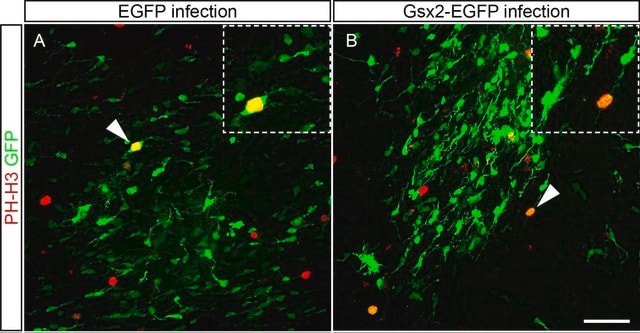
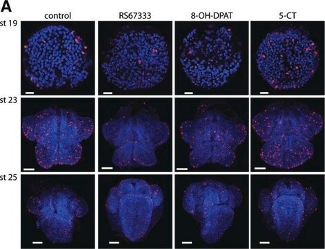
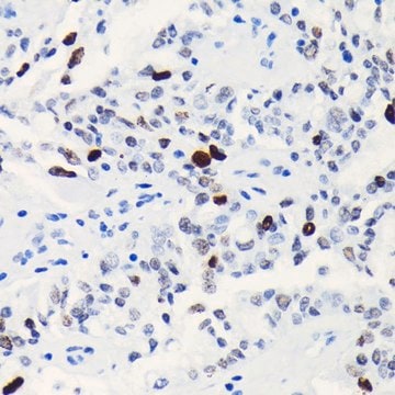
Anti-phospho-Histone H3 (pSer10) antibody produced in rabbit has been used in the detection of pSer10-histone H3 by immunoblotting, immunohistochemistry and by immunofluorescence.
Rabbit polyclonal anti-phospho-Histone H3 (pSer10) antibody is used to tag Ser10 phosphorylated histone H3 for detection and quantitation by immunocytochemical and immunohistochemical (IHC) techniques such as immunoblotting and immunofluorescence. It is used as a probe to determine the presence and roles of Ser10 phosphorylated histone H3 in chromosome condensation during both mitosis and meiosis.

Actions biochimiques/physiologiques

Phosphorylation of histone H3, referred to as the nucleosomal response, is localized to a small fraction of highly acetylated H3s and occurs primarily in response to mitogenic and stress stimuli. Histone H3 is phosphorylated during mitosis on at least two serine residues, Ser10 and Ser28. Phosphorylation of Ser10 is considered a crucial event for the onset of mitosis. It is tightly correlated with chromosome condensation during both mitosis and meiosis. Members of the Aurora/AIK family of protein kinases are involved in the mitotic phosphorylation of histone H3 at Ser10 residue. Expression of Aurora-A and Aurora-B is tightly coordinated with histone H3 phosphorylation at G2/M transition and both kinases efficiently phosphorylate H3 at Ser10 in vitro and in vivo. Phosphorylation of H3 at Ser10 is also directly correlated with the induction of immediate-early genes such as c-jun, c-fos and c-myc. Protein kinase A (PKA), ribosomal protein S6 kinase-3 (Rsk-2) and mitogen and stress activated protein kinase-1 (Msk-1) are necessary for the histone H3 phosphorylation.

Forme physique

Solution in 0.01 M phosphate buffered saline, pH 7.4, containing 15 mM sodium azide.

Clause de non-responsabilité

Unless otherwise stated in our catalog or other company documentation accompanying the product(s), our products are intended for research use only and are not to be used for any other purpose, which includes but is not limited to, unauthorized commercial uses, in vitro diagnostic uses, ex vivo or in vivo therapeutic uses or any type of consumption or application to humans or animals.

Vous ne trouvez pas le bon produit ?  

Essayez notre Outil de sélection de produits.

Code de la classe de stockage

12 - Non Combustible Liquids

Classe de danger pour l'eau (WGK)

nwg

Point d'éclair (°F)

Not applicable

Point d'éclair (°C)

Not applicable


Certificats d'analyse (COA)

Recherchez un Certificats d'analyse (COA) en saisissant le numéro de lot du produit. Les numéros de lot figurent sur l'étiquette du produit après les mots "Lot" ou "Batch".

Déjà en possession de ce produit ?

Retrouvez la documentation relative aux produits que vous avez récemment achetés dans la Bibliothèque de documents.

Consulter la Bibliothèque de documents

AMOTL1 promotes breast cancer progression and is antagonized by merlin
Couderc C, et al.
Neoplasia, 18(1), 10-24 (2016)
Phosphoacetylation of histone H3 on c-fos-and c-jun-associated nucleosomes upon gene activation
Clayton AL, et al.
The Embo Journal, 19(14), 3714-3726 (2000)
Mitotic phosphorylation of histone H3: spatio-temporal regulation by mammalian Aurora kinases
Crosio C, et al.
Molecular and Cellular Biology, 22(3), 874-885 (2002)
Ashley M Driver et al.
Journal of developmental biology, 5(4) (2018-04-05)
Proper cerebellar development is dependent on tightly regulated proliferation, migration, and differentiation events. Disruptions in any of these leads to a range of cerebellar phenotypes from ataxia to childhood tumors. Animal models have shown that proper regulation of sonic hedgehog
Immunohistochemistry for histone H3G34W and H3K36M is highly specific for giant cell tumor of bone and chondroblastoma, respectively, in FNA and core needle biopsy
Schaefer IM, et al.
Cancer Cytopathology, 126(8), 552-566 (2018)

Notre équipe de scientifiques dispose d'une expérience dans tous les secteurs de la recherche, notamment en sciences de la vie, science des matériaux, synthèse chimique, chromatographie, analyse et dans de nombreux autres domaines..

Contacter notre Service technique