Accéder au contenu
Merck
Toutes les photos(1)

Key Documents

MABC1595

Sigma-Aldrich

Anti-RIPK3/RIP3 Antibody, clone 8G7

Synonyme(s) :

Anti-RIPK3 Antibody, Clone 8G7 Anti-RIPK3, RIPK3 Detection Antibody

Se connecterpour consulter vos tarifs contractuels et ceux de votre entreprise/organisme


About This Item

Code UNSPSC :
12352203
Nomenclature NACRES :
NA.43

Source biologique

rat

Niveau de qualité

Conjugué

unconjugated

Forme d'anticorps

purified antibody

Type de produit anticorps

primary antibodies

Clone

8G7, monoclonal

Poids mol.

calculated mol wt 53.32 kDa
observed mol wt ~53 kDa

Espèces réactives

mouse

Conditionnement

antibody small pack of 100 μL

Technique(s)

immunofluorescence: suitable
western blot: suitable

Isotype

IgG2aκ

Numéro d'accès UniProt

Conditions d'expédition

dry ice

Température de stockage

2-8°C

Modification post-traductionnelle de la cible

unmodified

Description générale

Receptor-interacting serine/threonine-protein kinase 3 (UniProt: EC:2.7.11.1, RIP-like protein kinase 3, Receptor-interacting protein 3, RIP-3, mRIP3) is encoded by the Ripk3 (also known as Rip3) gene (Gene ID: 56532) in murine species. RIPK3 is a serine/threonine protein kinase that is expressed in embryo and adult spleen, liver, testis, brain, and lung. Its protein kinase domain is localized in amino acids 22 to 293 in the N-terminal half and it also has a C-terminal RIP homolytic interaction motif (RHIM; aa 440-461), which mediates interaction with the RHIM motif of RIPK1 and facilitate the formation of a necrosome. RIPK3 is shown to activate both necroptosis and apoptosis. Activated RIPK3 forms a necrosis-inducing complex and mediates phosphorylation of MLKL, promoting MLKL localization to the plasma membrane and execution of programmed necrosis that is characterized by calcium influx and plasma membrane damage. Its location is mainly cytoplasmic, however, following infection, it can also be detected in the nucleus. RIPK3 also regulates apoptosis that is dependent on RIPK1, FADD, and CASP8, and is independent of MLKL and RIPK3 kinase activity. It phosphorylates RIPK1 and then RIPK1 and RIPK3 undergo reciprocal auto- and trans-phosphorylation. In some cell types, RIPK3 is also reported to restrict viral replication by promoting cell death-independent responses. Clone 8G7 is raised against an extended kinase domain (residues 2-353) of mouse RIPK3 and can detect forms that may not be detected by antibodies directed towards the C-terminus of the full-length RIPK3. (Ref.: Samson, AL., et al. (2021). Cell Death Differ. https://doi.org/10.1038/s41418-021-00742-x; Petrie, EJ., et al. (2019). Cell Rep. 28(13); 3309-3319).

Spécificité

Clone 8G7 is a rat monoclonal antibody that detects murine Receptor-interacting serine/threonine-protein kinase 3 (RIPK3). It targets an epitope within the extended kinase domain that includes most of the N-terminal half and a part of C-terminal half.

Immunogène

Recombinant fragment corresponding to 352 amino acids from the extended kinase domain of murine Receptor-interacting serine/threonine-protein kinase 3 (RIPK3).

Application

Quality Control Testing

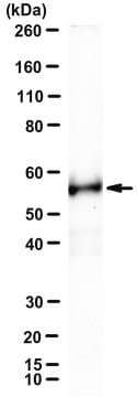
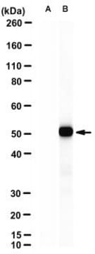
Evaluated by Western Blotting in Mouse dermal fibroblast lysates.

Western Blotting Analysis: A 1:500 dilution of this antibody detected RIPK3/RIP3 in Mouse dermal fibroblast lysates.

Tested Applications

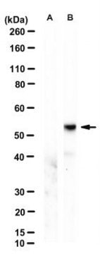
Western Blotting Analysis: A representative lot detected RIPK3/RIP3 in Western Blotting applications (Petrie, E.J., et. al. (2019). Cell Rep. 28(13):3309-3319).

Immunofluorescence Analysis: A representative lot detected RIPK3/RIP3 in Immunofluorescence applications (Samson, A.L., et. al. (2021). Cell Death Differ. doi: 10.1038/s41418-021-00742-x)

Note: Actual optimal working dilutions must be determined by end user as specimens, and experimental conditions may vary with the end user
Anti-RIPK3/RIP3, clone 8G7, Cat. No. MABC1595, is a rat monoclonal antibody that detects RIPK3/RIP3 and is tested for use in Immunofluorescence and Western Blotting.

Forme physique

Purified rat monoclonal antibody IgG2a in buffer containing 0.1 M Tris-Glycine (pH 7.4), 150 mM NaCl with 0.05% sodium azide.

Stockage et stabilité

Recommend storage at +2°C to +8°C. For long term storage antibodies can be kept at -20°C. Avoid repeated freeze-thaws.

Autres remarques

Concentration: Please refer to the Certificate of Analysis for the lot-specific concentration.

Clause de non-responsabilité

Unless otherwise stated in our catalog or other company documentation accompanying the product(s), our products are intended for research use only and are not to be used for any other purpose, which includes but is not limited to, unauthorized commercial uses, in vitro diagnostic uses, ex vivo or in vivo therapeutic uses or any type of consumption or application to humans or animals.

Vous ne trouvez pas le bon produit ?  

Essayez notre Outil de sélection de produits.

Code de la classe de stockage

12 - Non Combustible Liquids

Classe de danger pour l'eau (WGK)

WGK 1

Point d'éclair (°F)

Not applicable

Point d'éclair (°C)

Not applicable


Certificats d'analyse (COA)

Recherchez un Certificats d'analyse (COA) en saisissant le numéro de lot du produit. Les numéros de lot figurent sur l'étiquette du produit après les mots "Lot" ou "Batch".

Déjà en possession de ce produit ?

Retrouvez la documentation relative aux produits que vous avez récemment achetés dans la Bibliothèque de documents.

Consulter la Bibliothèque de documents

Notre équipe de scientifiques dispose d'une expérience dans tous les secteurs de la recherche, notamment en sciences de la vie, science des matériaux, synthèse chimique, chromatographie, analyse et dans de nombreux autres domaines..

Contacter notre Service technique