Accéder au contenu
Merck
Toutes les photos(3)

Principaux documents

AB10554

Sigma-Aldrich

Anti-Tbr1 Antibody

from rabbit, purified by affinity chromatography

Synonyme(s) :

T-box, brain, 1, T-brain-1, T-box brain protein 1

Se connecterpour consulter vos tarifs contractuels et ceux de votre entreprise/organisme


About This Item

Code UNSPSC :
12352203
eCl@ss :
32160702
Nomenclature NACRES :
NA.41

Source biologique

rabbit

Niveau de qualité

Forme d'anticorps

affinity isolated antibody

Type de produit anticorps

primary antibodies

Clone

polyclonal

Produit purifié par

affinity chromatography

Espèces réactives

mouse

Réactivité de l'espèce (prédite par homologie)

canine (based on 100% sequence homology), canine, bovine, opossum, horse, rat, pig

Technique(s)

immunohistochemistry: suitable (paraffin)
western blot: suitable

Numéro d'accès NCBI

Numéro d'accès UniProt

Conditions d'expédition

wet ice

Modification post-traductionnelle de la cible

unmodified

Informations sur le gène

human ... TBR1(10716)

Description générale

T-box brain protein 1 (UniProt: Q64336; also known as T-brain-1, TBR-1, TES-56) is encoded by the Tbr1 gene (Gene ID: 21375) in murine species. Tbr1 is a homodimeric nuclear protein that serves as a transcriptional repressor and is involved in multiple aspects of cortical development, including neuronal migration, laminar and areal identity, and axonal projection. As transcriptional repressor of FEZ Family Zinc Finger 2 (FEZF2), it blocks the formation of the corticospinal (CS) tract from layer 6 projection neurons, thereby restricting the origin of CS axons specifically to layer 5 neurons. Its T-box DNA binding domain is localized in amino acids 213-393. Its expression is observed in developing and adult cerebral cortex and in olfactory bulbs. It is detected around day 10 of embryonic development in the preplate and at day 12.5 in the cortical plate. It is highly expressed in glutamatergic early-born cortical neurons. Tbr1 null mice exhibit severe defects of frontal cortex and layer 6 differentiation. (Ref.: Han, W., et al. (2011). Proc. Natl. Acad. Sci. USA. 108(7); 3041-3046; Bedogni, F., et al (2010). Proc. Natl Acad. Sci. USA. 107(29); 13129-13134; Hevner, RF., et al. (2001). Neuron. 29(2); 353-366).

Spécificité

This rabbit polyclonal antibody detects T-box brain protein 1 (Tbr1).

Immunogène

KLH-conjugated linear peptide corresponding to 18 amino acids from the N-terminal region of mouse T-box brain protein 1 (Tbr1).
KLH-conjugated linear peptide corresponding to mouse Tbr1 near the N-terminus.

Application

Quality Control Testing

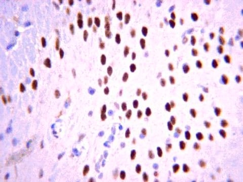
Evaluated by Immunohistochemistry (Paraffin) in Mouse brain tissue sections.Immunohistochemistry (Paraffin) Analysis: A 1:400 dilution of this antibody detected Tbr1 in Mouse cerebral cortex and cerebellum tissue sections.


Tested Applications

Western Blotting Analysis: A 1:500 dilution from a representative lot detected Tbr1 in Mouse fetal brain tissue lysate.Note: Actual optimal working dilutions must be determined by end user as specimens, and experimental conditions may vary with the end user.

Qualité

Evaluated by Immunohistochemistry in mouse frontal cortex tissue.

Immunohistochemistry Analysis: 1:400 dilution of this antibody detected Tbr1 in mouse frontal cortex tissue.

Description de la cible

~76 kDa observed; 73.94 kDa calculated. Uncharacterized bands may be observed in some lysate(s).

Forme physique

Affinity purified
Purified rabbit polyclonal antibody in buffer containing 0.1 M Tris-Glycine (pH 7.4), 150 mM NaCl with 0.05% sodium azide.

Stockage et stabilité

Recommended storage: +2°C to +8°C.

Remarque sur l'analyse

Control
Mouse frontal cortex tissue

Autres remarques

Concentration: Please refer to the Certificate of Analysis for the lot-specific concentration.

Clause de non-responsabilité

Unless otherwise stated in our catalog or other company documentation accompanying the product(s), our products are intended for research use only and are not to be used for any other purpose, which includes but is not limited to, unauthorized commercial uses, in vitro diagnostic uses, ex vivo or in vivo therapeutic uses or any type of consumption or application to humans or animals.

Vous ne trouvez pas le bon produit ?  

Essayez notre Outil de sélection de produits.

Code de la classe de stockage

12 - Non Combustible Liquids

Classe de danger pour l'eau (WGK)

WGK 1

Point d'éclair (°F)

Not applicable

Point d'éclair (°C)

Not applicable


Certificats d'analyse (COA)

Recherchez un Certificats d'analyse (COA) en saisissant le numéro de lot du produit. Les numéros de lot figurent sur l'étiquette du produit après les mots "Lot" ou "Batch".

Déjà en possession de ce produit ?

Retrouvez la documentation relative aux produits que vous avez récemment achetés dans la Bibliothèque de documents.

Consulter la Bibliothèque de documents

Yuko Muroyama et al.
PLoS genetics, 12(12), e1006514-e1006514 (2016-12-28)
Mitral cells (MCs) of the mammalian olfactory bulb have a single primary dendrite extending into a single glomerulus, where they receive odor information from olfactory sensory neurons (OSNs). Molecular mechanisms for controlling dendritic arbors of MCs, which dynamically change during
Martina Mavrovic et al.
EMBO reports, 21(4), e48880-e48880 (2020-02-18)
KCC2, encoded in humans by the SLC12A5 gene, is a multifunctional neuron-specific protein initially identified as the chloride (Cl- ) extruder critical for hyperpolarizing GABAA receptor currents. Independently of its canonical function as a K-Cl cotransporter, KCC2 regulates the actin
Xue Li et al.
Cerebral cortex (New York, N.Y. : 1991), 30(7), 3960-3976 (2020-02-03)
De novo microdeletion of chromosome 2p15-16.1 presents clinically recognizable phenotypes that include mental retardation, autism, and microcephaly. Chromosomal maintenance 1 (CRM1) is a gene commonly missing in patients with 2p15-16.1 microdeletion and one of two genes found in the smallest
Christine Sauerland et al.
Cerebral cortex (New York, N.Y. : 1991), 28(1), 145-157 (2017-12-19)
A hallmark of mammalian brain evolution is the emergence of the neocortex, which has expanded in all mammalian infraclasses (Eutheria, Marsupialia, Monotremata). In eutherians, neocortical neurons derive from distinct neural stem and progenitor cells (NPCs). However, precise data on the
Jing-Qiong Kang et al.
Nature neuroscience, 18(7), 988-996 (2015-05-26)
Genetic epilepsy and neurodegenerative diseases are two common neurological disorders that are conventionally viewed as being unrelated. A subset of patients with severe genetic epilepsies who have impaired development and often go on to die of their disease respond poorly

Notre équipe de scientifiques dispose d'une expérience dans tous les secteurs de la recherche, notamment en sciences de la vie, science des matériaux, synthèse chimique, chromatographie, analyse et dans de nombreux autres domaines..

Contacter notre Service technique