71074
BetaBlue Staining Kit
Convenient visualization of β-gal in cells or tissues
Se connecterpour consulter vos tarifs contractuels et ceux de votre entreprise/organisme
About This Item
Code UNSPSC :
41106513
Nomenclature NACRES :
NA.31
Produits recommandés
Fabricant/nom de marque
Novagen®
Conditions de stockage
OK to freeze
Conditions d'expédition
wet ice
Description générale

Convenient visualization of β-gal in cells or tissues
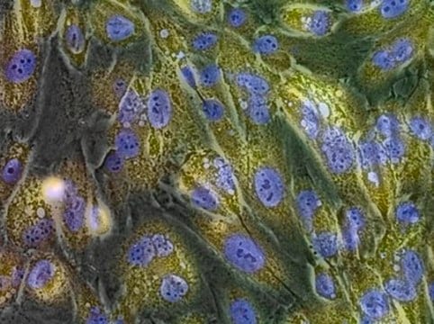
The BetaBlue Staining Kit provides direct visualization of β-galactosidase reporter expression in isolated cells, tissues, or intact organisms. The kit contains sufficient reagents for 100 staining reactions and includes solutions of the substrate X-Gal (5-bromo-4-chloro-3-indolyl-β-D-galactopyranoside) and Reaction Buffer optimized for rapid, sensitive histochemical staining with minimal background. The exceptional staining seen with the BetaBlue Staining Kit enables quick, accurate determination of transfection efficiencies, assessment of stable cell line generation, and transgene expression in tissue slices or whole mounts of transgenic animals.
Composants
•2 × 50 mlBetaBlue Reaction Buffer
•3 × 1 mlBetaBlue X-gal Solution
•3 × 1 mlBetaBlue X-gal Solution
Avertissement
Toxicity: Multiple Toxicity Values, refer to MSDS (O)
Informations légales
NOVAGEN is a registered trademark of Merck KGaA, Darmstadt, Germany
Code de la classe de stockage
10 - Combustible liquids
Certificats d'analyse (COA)
Recherchez un Certificats d'analyse (COA) en saisissant le numéro de lot du produit. Les numéros de lot figurent sur l'étiquette du produit après les mots "Lot" ou "Batch".
Déjà en possession de ce produit ?
Retrouvez la documentation relative aux produits que vous avez récemment achetés dans la Bibliothèque de documents.
Sebastian Frische et al.
American journal of physiology. Renal physiology, 320(1), F74-F86 (2020-12-08)
Variations in the claudin-14 (CLDN14) gene have been linked to increased risk of hypercalciuria and kidney stone formation. However, the exact cellular localization of CLDN14 and its regulation remain to be fully delineated. To this end, we generated a novel
Keishi Otsu et al.
Journal of bone and mineral research : the official journal of the American Society for Bone and Mineral Research, 31(11), 1943-1954 (2016-10-27)
During tooth development, oral epithelial cells differentiate into ameloblasts in order to form the most mineralized tissue in the vertebrate body: enamel. During this process, ameloblasts directionally secrete enamel matrix proteins and morphologically change from low columnar cells to polarized
Notre équipe de scientifiques dispose d'une expérience dans tous les secteurs de la recherche, notamment en sciences de la vie, science des matériaux, synthèse chimique, chromatographie, analyse et dans de nombreux autres domaines..
Contacter notre Service technique


