Pular para o conteúdo
Merck
Todas as fotos(3)

Documentos Principais

SAB4200867

Sigma-Aldrich

Anti-PMEL antibody produced in rabbit

affinity isolated antibody, buffered aqueous solution

Sinônimo(s):

ME20-M (ME20M) Melanoma-associated ME20 antigen, Melanocyte protein, Melanocyte protein Pmel 17, Melanocytes lineage-specific antigen GP100, Melanoma gp100, P1, P100, Premelanosome protein, Silver locus protein homolog

Faça loginpara ver os preços organizacionais e de contrato

Selecione um tamanho

100 μL
R$ 1.244,00
100 μL
R$ 3.156,00

R$ 1.244,00


Check Cart for Availability

Solicite uma grande encomenda

Selecione um tamanho

Alterar visualização
100 μL
R$ 1.244,00
100 μL
R$ 3.156,00

About This Item

Código UNSPSC:
12352203
NACRES:
NA.43

R$ 1.244,00


Check Cart for Availability

Solicite uma grande encomenda

forma do anticorpo

affinity isolated antibody

Nível de qualidade

Formulário

liquid

reatividade de espécies

human

concentração

~1 mg/mL

técnica(s)

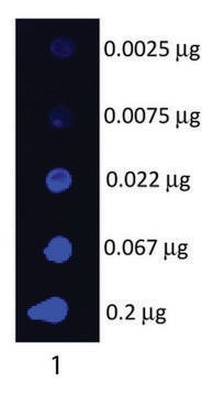
immunoblotting: 1:1000-1:2000 using human melanoma SK-MEL-28 cell lysate

nº de adesão UniProt

Condições de expedição

dry ice

temperatura de armazenamento

−20°C

modificação pós-traducional do alvo

unmodified

Descrição geral

The melanocyte-specific protein PMEL (also known as Pmel17, gp100, melanocyte protein, premelanosome protein or silver locus protein homolog (SILV)) is a type I transmembrane glycoprotein that is expressed primarily in pigment cells of the skin and eye. In melanosomes, the protein forms a fibrillar matrix on which the UV-shielding pigment melanin is deposited.1-2

Especificidade

Anti-PMEL antibody specifically recognizes human PMEL protein.

Aplicação

The antibody may be used in various immunochemical techniques including Immunoblotting (~100 kDa), Immunohistochemistry and Immunofluorescence.
Detection of the PMEL band by Immunoblotting is specifically inhibited by the immunogen.

Ações bioquímicas/fisiológicas

PMEL fibrils are a major functional component of the melanosomal compartment as they optimize melanin polymerization, condensation and storage.1,3
PMEL fibrils have an amyloidogenic nature and share features with pathological amyloids.4 Mutations in PMEL are associated with pigmentation disorders and/or impairments in eye development in various species.1,5,6
PMEL is suggested an excellent model system to study mechanisms of intracellular amyloid formation.1

forma física

Supplied as a solution in 0.01 M phosphate buffered saline pH 7.4, containing 15 mM sodium azide as a preservative.

Armazenamento e estabilidade

For continuous use, store at 2-8°C for up to one month. For extended storage, freeze in working aliquots. Repeated freezing and thawing is not recommended. If slight turbidity occurs upon prolonged storage, clarify the solution by centrifugation before use. Working dilution samples should be discarded if not used within 12 hours.

Exoneração de responsabilidade

Unless otherwise stated in our catalog  our products are intended for research use only and are not to be used for any other purpose, which includes but is not limited to, unauthorized commercial uses, in vitro diagnostic uses, ex vivo or in vivo therapeutic uses or any type of consumption or application to humans or animals.

Código de classe de armazenamento

12 - Non Combustible Liquids

Classe de risco de água (WGK)

WGK 1

Ponto de fulgor (°F)

Not applicable

Ponto de fulgor (°C)

Not applicable


Escolha uma das versões mais recentes:

Certificados de análise (COA)

Lot/Batch Number

Não está vendo a versão correta?

Se precisar de uma versão específica, você pode procurar um certificado específico pelo número do lote ou da remessa.

Já possui este produto?

Encontre a documentação dos produtos que você adquiriu recentemente na biblioteca de documentos.

Visite a Biblioteca de Documentos

Douglas M Fowler et al.
PLoS biology, 4(1), e6-e6 (2005-11-23)
Amyloid is a generally insoluble, fibrous cross-beta sheet protein aggregate. The process of amyloidogenesis is associated with a variety of neurodegenerative diseases including Alzheimer, Parkinson, and Huntington disease. We report the discovery of an unprecedented functional mammalian amyloid structure generated
Christin Bissig et al.
International journal of molecular sciences, 17(9) (2016-09-03)
In pigment cells, melanin synthesis takes place in specialized organelles, called melanosomes. The biogenesis and maturation of melanosomes is initiated by an unpigmented step that takes place prior to the initiation of melanin synthesis and leads to the formation of
Thomas Burgoyne et al.
Journal of cell science, 128(7), 1400-1407 (2015-02-19)
Analysis of melanosome biogenesis in the retinal pigment epithelium (RPE) is challenging because it occurs predominantly in a short embryonic time window. Here, we show that the zebrafish provides an ideal model system for studying this process because in the
Helia B Schonthaler et al.
Developmental biology, 284(2), 421-436 (2005-07-19)
Forward genetic screens have been instrumental in defining molecular components of visual function. The zebrafish mutant fading vision (fdv) has been identified in such a screen due to defects in vision accompanied by hypopigmentation in the retinal pigment epithelium (RPE)
Jia Shee Hee et al.
Scientific reports, 7, 44064-44064 (2017-03-09)
PMEL is a pigment cell protein that forms physiological amyloid in melanosomes. Many amyloids and/or their oligomeric precursors are toxic, causing or contributing to severe, incurable diseases including Alzheimer's and prion diseases. Striking similarities in intracellular formation pathways between PMEL

Questions

Reviews

No rating value

Active Filters

Nossa equipe de cientistas tem experiência em todas as áreas de pesquisa, incluindo Life Sciences, ciência de materiais, síntese química, cromatografia, química analítica e muitas outras.

Entre em contato com a assistência técnica