Pular para o conteúdo
Merck
Todas as fotos(1)

Documentos Principais

MABN2438

Sigma-Aldrich

Anti-RPE65 Antibody, clone KPSA1

Sinônimo(s):

All-trans-retinyl-palmitate hydrolase, EC:3.1.1.64, Lutein isomerase, Meso-zeaxanthin isomerase, Retinal pigment epithelium-specific 65 kDa protein, Retinoid isomerohydrolase, Retinol isomerase

Faça loginpara ver os preços organizacionais e de contrato


About This Item

Código UNSPSC:
12352203
NACRES:
NA.41
Preço e disponibilidade não estão disponíveis no momento.

fonte biológica

mouse

Nível de qualidade

forma do anticorpo

purified antibody

tipo de produto de anticorpo

primary antibodies

clone

KPSA1, monoclonal

reatividade de espécies

bovine, human, mouse

embalagem

antibody small pack of 100

técnica(s)

direct ELISA: suitable
immunohistochemistry (formalin-fixed, paraffin-embedded sections): suitable
western blot: suitable

Isotipo

IgG1κ

sequência de epítopo

C-terminal

nº de adesão de ID de proteína

nº de adesão UniProt

Informações sobre genes

human ... RPE65(6121)

Especificidade

Clone KPSA1 is a mouse monoclonal antibody that detects RPE65. It targets an epitope within 23 amino acid from the C-terminal region.

Imunogênio

Recombinant full-length Human RPE65 expressed in Sf9 cells.

Aplicação

Quality Control Testing

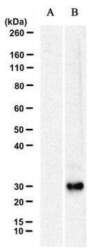
Evaluated by Western Blotting in Bovine retina microsomal preparation.

Western Blotting Analysis: A 1:10,000 dilution of this antibody detected RPE65 in Bovine retina microsomal preparation.

Tested Applications

Western Blotting Analysis: A representative lot detected RPE65 in Western Blotting applications (Golczak, M., et al. (2010). J Biol Chem. 285(13):9667-9682; Banskota, S., et al. (2022). Cell. 185(2):250-265.e16).

Immunoaffinity Purification: A representative lot was used for purification of crossed-linked RPE65.(Golczak, M., et al. (2010). J Biol Chem. 285(13):9667-9682).

Immunohistochemistry Applications: A representative lot detected RPE65 in Immunohistochemistry applications (Amengual, J., et al. (2014). Hum Mol Genet. 23(20):5402-17).

ELISA Analysis: A representative lot detected RPE65 in ELISA applications (Golczak, M., et al. (2010). J Biol Chem. 285(13):9667-9682).

Note: Actual optimal working dilutions must be determined by end user as specimens, and experimental conditions may vary with the end user.

Descrição-alvo

Retinoid isomerohydrolase (UniProt: Q16518; also known as EC:3.1.1.64, All-trans-retinyl-palmitate hydrolase, Lutein isomerase, Meso-zeaxanthin isomerase, Retinal pigment epithelium-specific 65 kDa protein, Retinol isomerase) is encoded by the RPE65 gene (Gene ID: 6121) in human. Retinol isomerase is a lipid-anchor protein that is attached to the membrane when palmitoylated and is present in soluble form when unpalmitoylated. It is shown to be critical for retinoid cycle and is involved in regeneration of 11-cis-retinal, the chromophore of rod and cone opsins. It catalyzes the cleavage and isomerization of all-trans-retinyl fatty acid esters to 11-cis-retinol that is further oxidized by 11-cis retinol dehydrogenase to 11-cis-retinal for use as visual chromophore. It can also catalyze the isomerization of lutein to meso-zeaxanthin an eye-specific carotenoid. The soluble form is reported to binds vitamin A (all-trans-retinol), making it available for lecithin retinol acyltransferase (LRAT) processing to all-trans-retinyl ester. The membrane form, palmitoylated by LRAT, binds all-trans-retinyl esters, making them available for isomerohydrolase processing to all-cis-retinol. The soluble form can be regenerated by transferring its palmitoyl groups onto 11-cis-retinol, a reaction catalyzed by LRAT. It undergoes light-dependent intracellular transport to become more concentrated in the central region of the retina pigment epithelium cells. Mutations in RPE65 gene are known to cause Retinitis pigmentosa that is characterized by retinal pigment deposits visible on fundus examination and primary loss of rod photoreceptor cells followed by secondary loss of cone photoreceptors. (Ref.: Golczak, M., et al. (2010). J. Biol. Chem. 285(13); 9667-9682; Moiseyev, G., et al. (2005). Proc. Natl. Acad. Sci. USA. 102(35):12413-12418).

forma física

Purified mouse monoclonal antibody IgG1 in buffer containing 0.1 M Tris-Glycine (pH 7.4), 150 mM NaCl and 0.05% sodium azide.

Reconstituição

1.0 mg/mL. Please refer to guidance on suggested starting dilutions and/or titers per application and sample type.

Armazenamento e estabilidade

Recommended storage: +2°C to +8°C.

Outras notas

Concentration: Please refer to the Certificate of Analysis for the lot-specific concentration.

Exoneração de responsabilidade

Unless otherwise stated in our catalog or other company documentation accompanying the product(s), our products are intended for research use only and are not to be used for any other purpose, which includes but is not limited to, unauthorized commercial uses, in vitro diagnostic uses, ex vivo or in vivo therapeutic uses or any type of consumption or application to humans or animals.

Não está encontrando o produto certo?  

Experimente o nosso Ferramenta de seleção de produtos.

Código de classe de armazenamento

12 - Non Combustible Liquids

Classe de risco de água (WGK)

WGK 1

Ponto de fulgor (°F)

Not applicable

Ponto de fulgor (°C)

Not applicable


Certificados de análise (COA)

Busque Certificados de análise (COA) digitando o Número do Lote do produto. Os números de lote e remessa podem ser encontrados no rótulo de um produto após a palavra “Lot” ou “Batch”.

Já possui este produto?

Encontre a documentação dos produtos que você adquiriu recentemente na biblioteca de documentos.

Visite a Biblioteca de Documentos

Questions

Reviews

No rating value

Active Filters

Nossa equipe de cientistas tem experiência em todas as áreas de pesquisa, incluindo Life Sciences, ciência de materiais, síntese química, cromatografia, química analítica e muitas outras.

Entre em contato com a assistência técnica