Pular para o conteúdo
Merck
Todas as fotos(2)

Documentos Principais

MABC604

Sigma-Aldrich

Anti-MLKL Antibody, clone 3H1

clone 3H1, from rat

Sinônimo(s):

Mixed lineage kinase domain-like protein, MLKL

Faça loginpara ver os preços organizacionais e de contrato


About This Item

Código UNSPSC:
12352203
eCl@ss:
32160702
NACRES:
NA.41
Preço e disponibilidade não estão disponíveis no momento.

fonte biológica

rat

Nível de qualidade

forma do anticorpo

purified antibody

tipo de produto de anticorpo

primary antibodies

clone

3H1, monoclonal

reatividade de espécies

mouse

técnica(s)

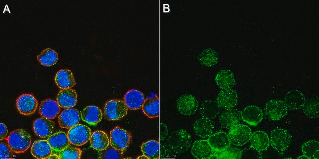
immunocytochemistry: suitable
immunoprecipitation (IP): suitable
western blot: suitable

Isotipo

IgG

nº de adesão NCBI

nº de adesão UniProt

Condições de expedição

wet ice

modificação pós-traducional do alvo

unmodified

Informações sobre genes

mouse ... Mlkl(74568)

Descrição geral

MLKL, also known as Mixed lineage kinase domain-like protein, and encoded by the gene MLKL, is an interesting protein that is required for the execution of programmed necrosis or necroptosis, for example in response to TNF-alpha induced necrosis. MLKL is a component of the "necrosome," the multiprotein complex that triggers tumor necrosis factor (TNF)-induced cell death by necroptosis. The programmed necrosis induced by TNF-α also requires the activities of the receptor-interacting serine-threonine kinases RIP1 and RIP3 and they too interact directly with the mixed lineage kinase domain-like protein MLKL. RIP3 is an essential upstream kinase in necroptosis. The pseudokinase MLKL functions as a substrate of RIP3 to mediate downstream signaling and when complexed to MLKL the RIP3-MLKL complex is stabilized by AMP-PNP to adopt an inactive conformation.

Imunogênio

Epitope: Brace region.
KLH-conjugated linear peptide corresponding to a sequence from the two-helix linker (Brace) region N-terminal to the pseudokinase domain of mouse MLKL (Hildebrand, J.M., et al. (2014). Proc. Natl. Acad. Sci. U.S.A. 111(42):15072-15077; Murphy, J.M., et al. (2013). Immunity. 39(3):443-453).

Aplicação

Immunocytochemistry Analysis: A representative lot detected MLKL in mouse dermal fibroblasts (MDFs) from wild-type, but not Mlkl-/- mice (Courtesy of Prof. James M. Murphy, Walter and Eliza Hall Institute, Australia).

Immunoprecipitation Analysis: A representative lot immunoprecipitated MLKL from soluble extract of Mlkl-/- mouse dermal fibroblasts (MDFs) harboring doxycycline-inducible wild-type mouse MLKL expression construct only after, but not before doxycycline treatment (Murphy, J.M., et al. (2013). Immunity. 39(3):443-453).

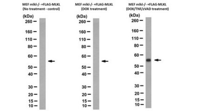
Western Blotting Analysis: A representative lot detected Q-VD-OPh (TSQ; Cat. No. 551476) treatment-induced membrane translocation of murine and equine MLKL N-terminal fragment (a.a. 1-180 and 1-189, respectively) exogenously expressed in mouse dermal fibroblasts (MDFs) from Mlkl-/- mice (Tanzer, M.C., et al. (2016). Cell Death Differ.. In press).

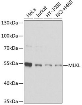
Western Blotting Analysis: A representative lot detected MLKL in human HT-29 and U937 cells (Tanzer, M.C., et al. (2015). Biochem. J. 471(2):255-265).

Western Blotting Analysis: Representative lots detected MLKL membrane translocation in mouse dermal fibroblasts (MDFs) upon Q-VD-OPh (TSQ; Cat. No. 551476) treatment (Tanzer, M.C., et al. (2015). Biochem. J. 471(2):255-265; Hildebrand, J.M., et al. (2014). Proc. Natl. Acad. Sci. U.S.A. 111(42):15072-15077).

Western Blotting Analysis: Representative lots detected MLKL expression in L292 mouse fibroblasts, as well as in mouse dermal fibroblasts (MDFs), mouse embryonic fibroblasts (MEFs) and bone marrow derived macrophages (BMDMs) from wild-type, but not Mlkl-/- mice (Cook, W.D., et al. (2014). Cell Death Differ. 21(10):1600-1612; Murphy, J.M., et al. (2013). Immunity. 39(3):443-453).

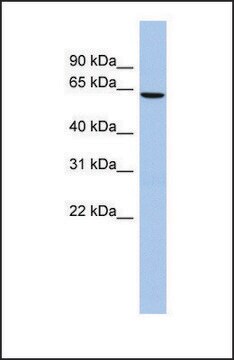
Western Blotting Analysis: A representative lot detected recombinant full-length mouse MLKL as well as a.a. 1-180 and a.a. 124-464, but not a.a. 1-125 or a.a. 179-464, MLKL fragments (Hildebrand, J.M., et al. (2014). Proc. Natl. Acad. Sci. U.S.A. 111(42):15072-15077).

Western Blotting Analysis: A representative lot detected MLKL expression in all tissues tested except brain from wild-type mice, while no target band was seen in any tissues from Mlkl-/- mice (Murphy, J.M., et al. (2013). Immunity. 39(3):443-453).
This Anti-MLKL Antibody, clone 3H1 is validated for use in Immunocytochemistry, Immunoprecipitation, and Western Blotting for the detection of MLKL.

Qualidade

Evaluated by Western Blotting in mouse heart tissue lysate.

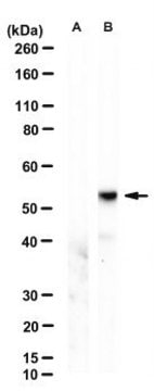
Western Blotting Analysis: 0.5 µg/mL of this antibody detected MLKL in 10 µg of mouse heart tissue lysate.

Descrição-alvo

~52 kDa observed. 54.48/30.28 (human isoform 1/2) and 54.32/53.38 kDa (mouse isoform 1/2) calculated. Uncharacterized band(s) may be observed in some cell lysates.

forma física

Format: Purified
Purified rat IgG in buffer containing 0.1 M Tris-Glycine (pH 7.4), 150 mM NaCl with 0.05% sodium azide.

Outras notas

Concentration: Please refer to the Certificate of Analysis for the lot-specific concentration.

Não está encontrando o produto certo?  

Experimente o nosso Ferramenta de seleção de produtos.

Código de classe de armazenamento

12 - Non Combustible Liquids

Classe de risco de água (WGK)

WGK 1

Ponto de fulgor (°F)

Not applicable

Ponto de fulgor (°C)

Not applicable


Certificados de análise (COA)

Busque Certificados de análise (COA) digitando o Número do Lote do produto. Os números de lote e remessa podem ser encontrados no rótulo de um produto após a palavra “Lot” ou “Batch”.

Já possui este produto?

Encontre a documentação dos produtos que você adquiriu recentemente na biblioteca de documentos.

Visite a Biblioteca de Documentos

RIPK1- and RIPK3-induced cell death mode is determined by target availability.
Cook, WD; Moujalled, DM; Ralph, TJ; Lock, P; Young, SN; Murphy, JM; Vaux, DL
Cell Death and Differentiation null
D M Moujalled et al.
Cell death & disease, 5, e1086-e1086 (2014-03-01)
Necroptosis is a mechanism by which cells can kill themselves that does not require caspase activity or the presence of the pro-apoptotic Bcl-2 family members Bax or Bak. It has been reported that RIPK3 (receptor interacting protein kinase 3) activates
A V Jacobsen et al.
Cell death & disease, 7, e2051-e2051 (2016-01-19)
Necroptosis is a caspase-independent form of regulated cell death that has been implicated in the development of a range of inflammatory, autoimmune and neurodegenerative diseases. The pseudokinase, Mixed Lineage Kinase Domain-Like (MLKL), is the most terminal known obligatory effector in
Joanne M Hildebrand et al.
Proceedings of the National Academy of Sciences of the United States of America, 111(42), 15072-15077 (2014-10-08)
Necroptosis is considered to be complementary to the classical caspase-dependent programmed cell death pathway, apoptosis. The pseudokinase Mixed Lineage Kinase Domain-Like (MLKL) is an essential effector protein in the necroptotic cell death pathway downstream of the protein kinase Receptor Interacting
M C Tanzer et al.
Cell death and differentiation, 23(7), 1185-1197 (2016-02-13)
The pseudokinase, MLKL (mixed-lineage kinase domain-like), is the most terminal obligatory component of the necroptosis cell death pathway known. Phosphorylation of the MLKL pseudokinase domain by the protein kinase, receptor interacting protein kinase-3 (RIPK3), is known to be the key

Questions

Reviews

No rating value

Active Filters

Nossa equipe de cientistas tem experiência em todas as áreas de pesquisa, incluindo Life Sciences, ciência de materiais, síntese química, cromatografia, química analítica e muitas outras.

Entre em contato com a assistência técnica