MABN1794
Anti-GnRH-R Antibody, clone F1G4
clone FIG4, from mouse
Sinónimos:
Gonadotropin-releasing hormone receptor, GnRH receptor, GnRH-R, Leutinizing hormone-releasing hormone receptor, Luteinizing-releasing hormone receptor
About This Item
Productos recomendados
biological source
mouse
Quality Level
antibody form
purified immunoglobulin
antibody product type
primary antibodies
clone
FIG4, monoclonal
species reactivity
human
technique(s)
ELISA: suitable
dot blot: suitable
flow cytometry: suitable
immunohistochemistry: suitable (paraffin)
western blot: suitable
isotype
IgG1λ
NCBI accession no.
UniProt accession no.
shipped in
wet ice
target post-translational modification
unmodified
Gene Information
human ... GNRHR (2798)
General description
Specificity
Immunogen
Application
ELISA Analysis: A representative lot detected the immobilized immunogen peptide by ELISA (Karande, A.A., et al. (1995). Mol. Cell. Endocrinol. 114(102):51-56).
Flow Cytometry Analysis: A representative lot detected surface GnRH-R immunoreactivity on live human breast carcinoma T47D and ovarian carcinoma OVCAR-3 cells. Most likely due to low affinity antibody binding or unoptimized antibody concentration used, only ~50% of the T47D and ~10% of the OVCAR-3 populations were stained (Karande, A.A., et al. (1995). Mol. Cell. Endocrinol. 114(102):51-56).
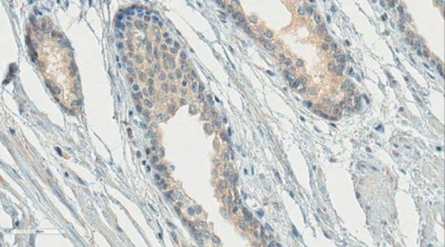
Immunohistochemistry Analysis: A rerepsentative lot detected GnRH-R expression on gonadotropin-producing endocrine cells (gonadotropes) in pituitary, as well as among neurons in human hippocampus and neocortex, using formalin-fixed, paraffin-embedded human tissue sections (Wilson, A.C., et al. (2006). J. Endocrinol. 191(3):651–663).
Immunohistochemistry Analysis: A rerepsentative lot detected similar hippocampus GnRH-R immunoreactivity among Alzheimer′s diseased (AD) and age-matched control brain sections, while significanly decreased GnRH-R immunoreactivity associated with the apical dendrites of pyramidal neurons was seen in the AD brain using formalin-fixed, paraffin-embedded tissue sections (Wilson, A.C., et al. (2006). J. Endocrinol. 191(3):651–663).
Immunohistochemistry Analysis: A rerepsentative lot detected GnRH-R-positive cells in the anterior pituitary, but not the posterior pituitary using frozen human brain sections. Pre-blocking with immunogen peptide abolished the antibody staining (Karande, A.A., et al. (1995). Mol. Cell. Endocrinol. 114(102):51-56).
Western Blotting Analysis: A rerepsentative lot detected the expression of multiple GnRH-R variants in human cortex and pituitary tissue lysates, including the most prominent 30 kDa, 64 kDa, and 136 kDa bands. The 136 kDa band was seen significantly downregulated in the cortex, but not pituitary from Alzheimer′s diseased (AD) brain when compared to age-matched control (Wilson, A.C., et al. (2006). J. Endocrinol. 191(3):651–663).
Neuroscience
Hormones & Receptors
Quality
Western Blotting Analysis: 2.0 µg/mL of this antibody detected GnRH-R in 10 µg of human pituitary tissue lysate.
Target description
Physical form
Storage and Stability
Other Notes
Disclaimer
¿No encuentra el producto adecuado?
Pruebe nuestro Herramienta de selección de productos.
Storage Class
12 - Non Combustible Liquids
wgk_germany
WGK 1
flash_point_f
Not applicable
flash_point_c
Not applicable
Certificados de análisis (COA)
Busque Certificados de análisis (COA) introduciendo el número de lote del producto. Los números de lote se encuentran en la etiqueta del producto después de las palabras «Lot» o «Batch»
¿Ya tiene este producto?
Encuentre la documentación para los productos que ha comprado recientemente en la Biblioteca de documentos.
Nuestro equipo de científicos tiene experiencia en todas las áreas de investigación: Ciencias de la vida, Ciencia de los materiales, Síntesis química, Cromatografía, Analítica y muchas otras.
Póngase en contacto con el Servicio técnico