General description
Cadherin-5 (UniProt P33151; also known as 7B4 antigen, CD144, Vascular endothelial cadherin, VE-Cad, VE-cadherin) is encoded by the CDH5 gene (Gene ID 1003) in human. Cadherins (calcium-dependent adhesion) are type-1 transmembrane proteins that form adherens junctions in tissues and play important roles in mediating cell adhesion. Cadherins are designated with a prefix that specifies their tissue association. Vascular endothelial-cadherin (VE-cadherin) is the transmembrane component of the endothelial adherens junction between vascular endothelial cells (ECs) and plays a pivotal role in endothelium integrity and in the control of vascular permeability. One characteristic of VEGF-induced vascular permeability is the phosphorylation of VE-cadherin by Src family kinases (SFK), which leads to VE-cadherin internalization and the destabilization of adherens junctions. Likewise, VE-cadherin overexpression is shown to decrease the permeability of endothelial monolayers in vitro. SFK activation by dominant negative Csk overexpression is reported to result in VE-cadherin phosphorylation at tyrosines 658, 685, and 731 in human dermal microvascular endothelial cells (HDMECs) without affecting HDMEC monolayer permeability. On the other hand, expression of constitutively active Src promoted VE-cadherin phosphorylation on tyrosines 658 and 731 and decreased barrier function, suggesting that concurrent signaling events in addition to VE-cadherin tyrosine phosphorylation are needed to promote barrier permeability in response to inflammatory mediators or growth factors. VE-cadherin is initially produced with a signal peptide (a.a. 1-24) and a propeptide (a.a. 25-45) sequence, the removal of which yields tthe mature protein containing a large extracellular region (a.a. 46-599) with five cadherine repeats (a.a. 46-149, 150-256, 257-371, 372-476, 477-593), followed by a transmembrane segment (a.a. 600-620) and a cytoplasmic domain (a.a. 621-784).
Specificity
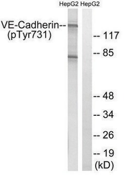
Target specificity of the unconjugated antibody (Cat. No. ABT1760) has been verified by immunogen peptide blocking in Western blotting application. Numbering of the target VE-cadherin phosphotyrosine residue varies depending on whether the N-terminal signal peptide sequence and propeptide sequence are included, as well as the species and spliced isoform a particular numbering is based on. Target pTyr this rabbit polyclonal detects corresponds to pTyr685/pTyr638 of human spliced isoform 1 precursor/mature sequence (UniProt P33151-1), pTyr570/pTyr523 of human spliced isoform 2 precursor/mature sequence (UniProt P33151-2), pTyr685/pTyr640 of bovine precursor/mature sequence (UniProt Q6URK6), and pTyr684/pTyr40 of porcine precursor/mature sequence (UniProt O02840). Equivalent target site sequence is not present in mouse, rat, or chicken VE-cadherin sequence.
Immunogen
Epitope: cytoplasmic domain
KLH-conjugated linear peptide corresponding to a sequence surrounding phosphorylated Tyr685 of human VE-cadherin isoform 1 precursor sequence.
Application
Research Category
Cell Structure
The unconjugated antibody (Cat. No. ABT1760) and Alexa Fluor™ 488-conjugated antibody (Cat. No. ABT1760-AF488) are also available for immunocytochemistry and Western blotting applications.
This rabbit polyclonal Anti-phospho-VE-Cadherin (Tyr685), Alexa Fluor™ 555 Conjugate, Cat. No. ABT1760-AF555 is validated for use in Immunocytochemistry for the detection of VE-cadherin Tyr685 phosphorylation.
Quality
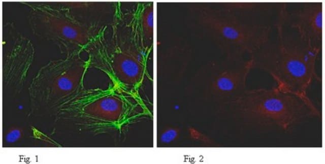
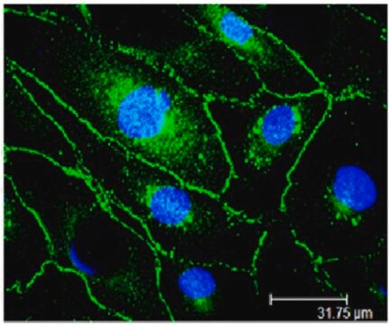
Evaluated by Immunocytochemistry in HUVECs.
Immunocytochemistry Analysis: A 1:100 dilution of this antibody detected a greatly upregulated VE-cadherin Tyr685 phosphorylation and internalization in serum-starved HUVECs upon VEGF-165 treatment (10 ng/mL for 10 min).
Target description
~130 kDa observed. Target band size appears larger than the calculated molecular weights of 82,58/69.55 kDa (human mature isoform 1/2), 87.53/74.50 kDa (human isoform 1/2 precursor), 82.80/87.47 kDa (bovine mature/precursor), and 82.45/87.55 kDa (porcine mature/precursor) due to glycosylation.
Physical form
Affinity purified.
Purified rabbit polyclonal antibody Alexa Fluor™ 555 conjugate in PBS with 1.5% BSA and 0.05% sodium azide.
Storage and Stability
Stable for 1 year at 2-8°C from date of receipt.
Other Notes
Concentration: Please refer to lot specific datasheet.
Legal Information
ALEXA FLUOR is a trademark of Life Technologies
Disclaimer
Unless otherwise stated in our catalog or other company documentation accompanying the product(s), our products are intended for research use only and are not to be used for any other purpose, which includes but is not limited to, unauthorized commercial uses, in vitro diagnostic uses, ex vivo or in vivo therapeutic uses or any type of consumption or application to humans or animals.


