General description
Gutamic acid decarboxylase (GAD; E.C. 4.1.1.15) is the enzyme responsible for the conversion of glutamic acid to gamma-aminobutyric acid (GABA), the major inhibitory transmitter in higher brain regions, and putative paracrine hormone in pancreatic islets. Two molecular forms of GAD (65 kDa and 67 kDa, 64% aa identity between forms) are highly conserved and both forms are expressed in the CNS, pancreatic islet cells, testis, oviduct and ovary. The isoforms are regionally distributed cytoplasmically in the brains of rats and mice (Sheikh, 1999). GAD65 is an amphiphilic, membrane-anchored protein (585 a.a.), encoded on human chromosome 10, and is responsible for vesicular GABA production. GAD67 is cytoplasmic (594 a.a.), encoded on chromosome 2, and seems to be responsible for significant cytoplasmic GABA production. GAD expression changes during neural development in rat spinal cord. GAD65 is expressed transiently in commissural axons around E13 but is down regulated the next day while GAD67 expression increases mostly in the somata of those neurons (Phelps, 1999). In mature rat pancreas, GAD65 and GAD67 appear to be differentially localized, GAD65 primarily in insulin-containing beta cells and GAD67 in glucagon-containing (A) cells (Li, 1995). GAD67 expression seems to be particularly plastic and can change in response to experimental manipulation (for example neuronal stimulation or transection) or disease progression and emergent disorders like schizophrenia (Volk, 2000). Colocalization of the two GAD isoforms also shows changes in GAD65/GAD67 distributions correlated with certain disease states such as IDDM and SMS.
Specificity
Anti-GAD67 Antibody, clone 1G10.2 reacts with the 67kDa isoform of Glutamate Decarboxylase (GAD67) of rat, mouse and human origins, other species not yet tested. No detectable cross-reactivity with GAD65 by Western blot on rat brain lysate when compared to blot probed with AB1511 that reacts with both GAD65 & GAD67.
Immunogen
Recombinant GAD67 protein
Application
Research Category
Neuroscience
Research Sub Category
Neurotransmitters & Receptors
Neuronal & Glial Markers
This Anti-GAD67 Antibody, clone 1G10.2 is validated for use in IH, IH(P), WB for the detection of GAD67. This Anti GAD antibody has more than 75 product citations.
Western Blot: A 1:5,000-1:10,000 dilution was used on a previous lot using ECL on rat and mouse brain lysate.
Immunohistochemistry on 4% paraformaldehyde fixed mouse and rat tissue: 1:1,000-1:5,000. The antibody has also worked well on human tissue fixed for 10 minutes in 4% PFA at 4°C or 100% ethanol at +20°C or 100% acetone at -20°C at dilutions of 1:2,500-1:5,000.
A polyclonal to GAD67 is also available as AB5992.
Optimal working dilutions must be determined by end user.
Quality
Routinely evaluated by immunohistochemistry by SKNSH cell lysate.
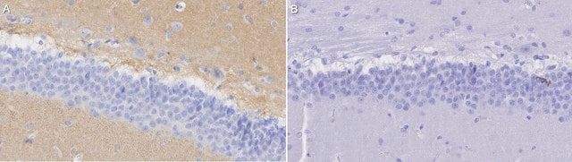
Immunohistochemistry(paraffin) Analysis:
Representative staining pattern and morphology of GAD67 in somatosensory 1, barrel field of the mouse cerebral cortex. All brown spots are Lamina VI a neurons. No Epitope retrieval was necessary. This lot of the antibody was diluted to 1:500, using IHC Select Detection with HRP-DAB.
Optimal Staining pattern/morphology of GAD67: Mouse Brain.
Linkage
Replaces: AB5992; AB5862P
Physical form
Format: Purified
Protein A purified
Purified mouse monoclonal IgG2a in buffer containing 0.1% sodium azide
Storage and Stability
Stable for 6 months at 2-8ºC in undiluted aliquots from date of receipt.
Analysis Note
Control
SKNSH cell lysate (human neuroblastoma), mouse cerebral cortex.
Legal Information
CHEMICON is a registered trademark of Merck KGaA, Darmstadt, Germany
Disclaimer
Unless otherwise stated in our catalog or other company documentation accompanying the product(s), our products are intended for research use only and are not to be used for any other purpose, which includes but is not limited to, unauthorized commercial uses, in vitro diagnostic uses, ex vivo or in vivo therapeutic uses or any type of consumption or application to humans or animals.