ABN78
Anti-NeuN Antibody (rabbit)
from rabbit, purified by affinity chromatography
Synonym(s):
RNA binding protein fox-1 homolog 3, Fox-1 homolog C, Fox-3, Hexaribonucleotide-binding protein 3, NeuN, Neuronal nuclei
About This Item
Recommended Products
biological source
rabbit
Quality Level
antibody form
affinity isolated antibody
antibody product type
primary antibodies
clone
polyclonal
purified by
affinity chromatography
species reactivity
mouse, human, snail, rat
technique(s)
immunocytochemistry: suitable
immunofluorescence: suitable
immunohistochemistry: suitable
western blot: suitable
NCBI accession no.
UniProt accession no.
shipped in
wet ice
storage temp.
2-8°C
target post-translational modification
unmodified
Gene Information
human ... RBFOX3(146713)
mouse ... Rbfox3(52897)
rat ... Rbfox3(287847)
General description
Specificity
Immunogen
Application
Immunohistochemistry Analysis: A 1:500 dilution from a representative lot detected NeuN-positive neurons in human (cerebellum and cerebral cortex) and mouse (hippocampus) brain tissue sections.
Immunocytochemistry Analysis: A representative lots immunostained 4% paraformaldehyde-fixed neurons isolated from snail brain (Nikolić, L., et al. (2013). J. Exp. Biol. 216(Pt 18):3531-3541).
Immunofluorescence Analysis: Representative lots detected NeuN-positive neurons in frozen brain sections from 4% paraformaldehyde-perfused mice (Cherry, J.D., et al. (2015). J. Neuroinflammation. 12:203; Huang, B., et al. (2015). Neuron. 85(6):1212-1226; Ataka, K., et al. (2013). PLoS One. 8(11):e81744).
Immunofluorescence Analysis: A representative lots detected NeuN-positive neurons in formalin-fixed and paraffin-embedded amyotrophic lateral sclerosis (ALS) human brain tissue sections (Fratta, P., et al. (2015). Neurobiol. Aging. 36(1):546.e1-7).
Immunohistochemistry Analysis: A representative lot detected NeuN-positive neurons in rat brain tissue sections (Mendonça, M.C., et al. (2013).Toxins. 5(12):2572-2588).
Western Blotting Analysis: Representative lot detected NeuN in mouse and rat brain tissue lysates (Huang, B., et al. (2015). Neuron. 85(6):1212-1226; Mendonça, M.C., et al. (2013).Toxins. 5(12):2572-2588).
This rabbit polyclonal antibody is also avaiable as Alexa Fluor™ 488 (ABN78A4), biotin (ABN78B), and Cy3 (ABN78C3) conjugates for immunocytochemistry, immunofluorescence, immunohistochemistry applications.
Neuroscience
Developmental Signaling
Quality
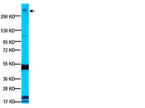
Western Blotting Analysis: 0.2 µg/mL of this antibody detected NeuN in 10 µg of mouse brain nuclear extract.
Target description
Physical form
Storage and Stability
Analysis Note
Mouse brain nuclear extract
Other Notes
Legal Information
Disclaimer
Not finding the right product?
Try our Product Selector Tool.
recommended
Storage Class Code
12 - Non Combustible Liquids
WGK
WGK 1
Flash Point(F)
Not applicable
Flash Point(C)
Not applicable
Certificates of Analysis (COA)
Search for Certificates of Analysis (COA) by entering the products Lot/Batch Number. Lot and Batch Numbers can be found on a product’s label following the words ‘Lot’ or ‘Batch’.
Already Own This Product?
Find documentation for the products that you have recently purchased in the Document Library.
Customers Also Viewed
Articles
Explore the basics of working with antibodies including technical information on structure, classes, and normal immunoglobulin ranges.
Explore the basics of working with antibodies including technical information on structure, classes, and normal immunoglobulin ranges.
Learn differences in monoclonal vs polyclonal antibodies including how antibodies are generated, clone numbers, and antibody formats.
Learn differences in monoclonal vs polyclonal antibodies including how antibodies are generated, clone numbers, and antibody formats.
Protocols
Tips and troubleshooting for FFPE and frozen tissue immunohistochemistry (IHC) protocols using both brightfield analysis of chromogenic detection and fluorescent microscopy.
Tips and troubleshooting for FFPE and frozen tissue immunohistochemistry (IHC) protocols using both brightfield analysis of chromogenic detection and fluorescent microscopy.
Tips and troubleshooting for FFPE and frozen tissue immunohistochemistry (IHC) protocols using both brightfield analysis of chromogenic detection and fluorescent microscopy.
Our team of scientists has experience in all areas of research including Life Science, Material Science, Chemical Synthesis, Chromatography, Analytical and many others.
Contact Technical Service


