
AB5320

Anti-CSPG4 Antibody
CHEMICON®, rabbit polyclonal
Synonym(s):
Chondroitin sulfate proteoglycan NG2, Melanoma chondroitin sulfate proteoglycan 3, Melanoma-associated chondroitin sulfate proteoglycan, chondroitin sulfate proteoglycan 4, chondroitin sulfate proteoglycan 4 (melanoma-associated)
Select a Size
| Pack Size | SKU | Availability | Price |
|---|---|---|---|
| 100 μg | Estimated to ship TODAY | $661.00 |
About This Item
$661.00
Estimated to ship TODAYDetails
Product Name
Anti-NG2 Chondroitin Sulfate Proteoglycan Antibody, Chemicon®, from rabbit
biological source
rabbit
Quality Segment
antibody form
purified antibody
antibody product type
primary antibodies
clone
polyclonal
purified by
affinity chromatography
species reactivity
rat, mouse, monkey, human
manufacturer/tradename
Chemicon®
technique(s)
ELISA: suitable, immunocytochemistry: suitable, immunohistochemistry: suitable, immunoprecipitation (IP): suitable, western blot: suitable
NCBI accession no.
UniProt accession no.
shipped in
dry ice
target post-translational modification
unmodified
Gene Information
human ... CSPG4(1464)
mouse ... Cspg4(121021)
rat ... Cspg4(81651)
rhesus monkey ... Cspg4(713086)
General description
Immunogen
Application
A 1:150-1:600 dilution of a previous lot was used in IC.
Immunohistochemistry:
1:200 dilution of a previous lot was used on frozen sections of embryonic mouse brain using an Alexa Fluor conjugated secondary antibody. Care should be taken to avoid overfixing tissue sections. A short (5-10 minute) fix in 2-4% PFA is recommended.
Immunoprecipitation:
2 μg/mL concentration of a previous lot was used in IP.
ELISA:
A 1:1,500-1:3,000 dilution of a previous lot was used in ELISA.
Optimal working dilutions and protocols must be determined by end user
Neuroscience
Neuronal & Glial Markers
Biochem/physiol Actions
Physical form
Preparation Note
Handling Recommendations:
Upon receipt, and prior to removing the cap, centrifuge the vial and gently mix the solution. Aliquot into microcentrifuge tubes and store at -20°C. Avoid repeated freeze/thaw cycles, which may damage IgG and affect product performance.
Analysis Note
Human melanoma, glioma and proliferating brain endothelial cells, rat B49 glial cell extracts or embryonic mouse brain tissue.
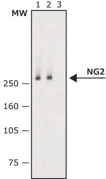
Western Blot: 1:1000 dilution of this lot detected NG2 chondroitin sulfate on 10 μg of rat brain lysates.
Other Notes
Legal Information
Disclaimer
1 of 1
This Item | |||
|---|---|---|---|
| biological source rabbit | biological source rabbit | biological source mouse | biological source mouse |
| antibody form purified antibody | antibody form purified antibody | antibody form purified immunoglobulin | antibody form purified from hybridoma cell culture, purified immunoglobulin |
| Quality Level 100 | Quality Level 100 | Quality Level 200 | Quality Level 200 |
| UniProt accession no. | UniProt accession no. | UniProt accession no. | UniProt accession no. - |
| clone polyclonal | clone polyclonal | clone 132.38, monoclonal | clone CS-56, monoclonal |
| technique(s) ELISA: suitable, immunohistochemistry: suitable, western blot: suitable, immunocytochemistry: suitable, immunoprecipitation (IP): suitable | technique(s) immunocytochemistry: suitable, immunohistochemistry: suitable | technique(s) immunocytochemistry: suitable, immunoprecipitation (IP): suitable, indirect ELISA: suitable, microarray: suitable, western blot: 1 μg/mL using rat brain neonatal extract | technique(s) immunofluorescence: 2.5-5 μg/mL using bovine BMGE cells, immunohistochemistry: 2.5-5 μg/mL using heat-retrieved formalin-fixed, paraffin-embedded human colon cancer sections and Biotin/ExtrAvidin ®-Peroxidase staining system. |



Still not finding the right product?
Explore all of our products under Anti-NG2 Chondroitin Sulfate Proteoglycan Antibody
— or —
Try our Product Selector Tool to narrow your options
Storage Class
12 - Non Combustible Liquids
wgk
WGK 2
flash_point_f
Not applicable
flash_point_c
Not applicable
Certificates of Analysis (COA)
Search for Certificates of Analysis (COA) by entering the products Lot/Batch Number. Lot and Batch Numbers can be found on a product’s label following the words ‘Lot’ or ‘Batch’.
Already Own This Product?
Find documentation for the products that you have recently purchased in the Document Library.
Protocols
Tips and troubleshooting for FFPE and frozen tissue immunohistochemistry (IHC) protocols using both brightfield analysis of chromogenic detection and fluorescent microscopy.
Articles
Antibodies combine with specific antigens to generate an exclusive antibody-antigen complex. Learn about the nature of this bond and its use as a molecular tag for research.
Immunofluorescence uses antibody-conjugated fluorescent molecules for protein localization, modification confirmation, and protein complex visualization.
Derivation and characterization of functional human neural stem cell derived oligodendrocyte progenitor cells (OPCs) that efficiently myelinate primary neurons in culture.
Related Content
Explore the basics of working with antibodies including technical information on structure, classes, and normal immunoglobulin ranges.
Learn differences in monoclonal vs polyclonal antibodies including how antibodies are generated, clone numbers, and antibody formats.
Global Trade Item Number
| SKU | GTIN |
|---|---|
| AB5320 | 04053252407413 |