推荐产品
生物源
mouse
品質等級
抗體表格
ascites fluid
抗體產品種類
primary antibodies
無性繁殖
PARV-19, monoclonal
物種活性
feline, rabbit, goat, frog, fish, human, pig, bovine, rat, mouse, canine
製造商/商標名
Chemicon®
技術
immunocytochemistry: suitable
immunohistochemistry: suitable (paraffin)
western blot: suitable
同型
IgG1
NCBI登錄號
UniProt登錄號
運輸包裝
dry ice
目標翻譯後修改
unmodified
基因資訊
human ... PVALB(5816)
一般說明
小清蛋白是一种钙结合清蛋白蛋白。 它具有三个EF手基序,并且在结构上与钙调蛋白和肌钙蛋白C有关。小清蛋白位于其水平最高的快速收缩肌肉以及大脑和一些内分泌组织中。 小清蛋白存在于神经的系统的GABA能中间神经元中,主要由皮质中的吊灯样细胞和篮状细胞表达。
特異性
小清蛋白。该抗体将与来自大脑和肌肉的小清蛋白反应。 通过免疫印迹,它识别12 kDa的蛋白。 该抗体针对第一个Ca+2-结合位点的表位,并特异性染色小清蛋白的Ca+2-结合形式。
免疫原
从青蛙肌肉中纯化的小清蛋白
應用
免疫组化:
间接免疫过氧化物酶以1:1,000-1:2,000的稀释度使用先前的批次。 该抗体对先前批次的福尔马林固定、石蜡包埋的大鼠小脑组织切片起作用。 固定似乎不会影响MAB1572’s检测小清蛋白的能力。
免疫细胞化学:
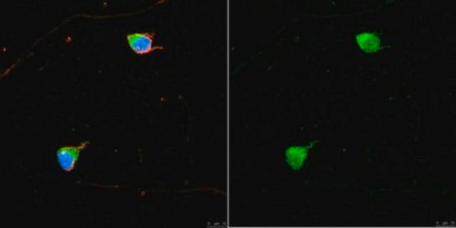
使用该抗体的先前批次用于培养神经元。
最佳的工作稀释度必须由最终用户确定。
间接免疫过氧化物酶以1:1,000-1:2,000的稀释度使用先前的批次。 该抗体对先前批次的福尔马林固定、石蜡包埋的大鼠小脑组织切片起作用。 固定似乎不会影响MAB1572’s检测小清蛋白的能力。
免疫细胞化学:
使用该抗体的先前批次用于培养神经元。
最佳的工作稀释度必须由最终用户确定。
抗小清蛋白抗体是用于 IH、IH(P)& WB的小清蛋白抗体。
研究子类别
信号神经科学
信号神经科学
研究类别
神经科学
神经科学
品質
通过蛋白质印迹对小鼠大脑裂解物进行常规评估。
蛋白质印迹分析:
该批次以1:1000的稀释度在10 μg小鼠脑裂解物中检测到小清蛋白。
蛋白质印迹分析:
该批次以1:1000的稀释度在10 μg小鼠脑裂解物中检测到小清蛋白。
標靶描述
12kda
外觀
未纯化
腹水小鼠单克隆IgG1液体
儲存和穩定性
自收到之日起在-20ºC可稳定保存1年。
分析報告
对照
脑组织,培养的神经元
脑组织,培养的神经元
其他說明
浓度:请参考批次特异性浓缩物的分析证书。
法律資訊
CHEMICON is a registered trademark of Merck KGaA, Darmstadt, Germany
免責聲明
除非我们的产品目录或产品附带的其他公司文档另有说明,否则我们的产品仅供研究使用,不得用于任何其他目的,包括但不限于未经授权的商业用途、体外诊断用途、离体或体内治疗用途或任何类型的消费或应用于人类或动物。
未找到合适的产品?
试试我们的产品选型工具.
儲存類別代碼
12 - Non Combustible Liquids
水污染物質分類(WGK)
nwg
閃點(°F)
Not applicable
閃點(°C)
Not applicable
其他客户在看
Mitochondrial calcium uptake underlies ROS generation during aminoglycoside-induced hair cell death.
Robert Esterberg et al.
The Journal of clinical investigation, 126(9), 3556-3566 (2016-08-09)
Exposure to aminoglycoside antibiotics can lead to the generation of toxic levels of reactive oxygen species (ROS) within mechanosensory hair cells of the inner ear that have been implicated in hearing and balance disorders. Better understanding of the origin of
Stress impairs GABAergic network function in the hippocampus by activating nongenomic glucocorticoid receptors and affecting the integrity of the parvalbumin-expressing neuronal network.
Hu, W; Zhang, M; Czeh, B; Flugge, G; Zhang, W
Neuropsychopharmacology null
Response features of parvalbumin-expressing interneurons suggest precise roles for subtypes of inhibition in visual cortex.
Runyan, CA; Schummers, J; Van Wart, A; Kuhlman, SJ; Wilson, NR; Huang, ZJ; Sur, M
Neuron null
Andrei Avanesov et al.
Methods in cell biology, 100, 153-204 (2010-11-30)
The zebrafish is one of the leading models for the analysis of the vertebrate visual system. A wide assortment of molecular, genetic, and cell biological approaches is available to study zebrafish visual system development and function. As new techniques become
Patricia R Jusuf et al.
The Journal of neuroscience : the official journal of the Society for Neuroscience, 31(7), 2549-2562 (2011-02-18)
Multipotent progenitors in the vertebrate retina often generate clonally related mixtures of excitatory and inhibitory neurons. The postmitotically expressed transcription factor, Ptf1a, is essential for all inhibitory fates in the zebrafish retina, including three types of horizontal and 28 types
我们的科学家团队拥有各种研究领域经验,包括生命科学、材料科学、化学合成、色谱、分析及许多其他领域.
联系技术服务部门


