
추천 제품


제품명
Monoclonal Anti-τ (Tau) antibody produced in mouse, clone TAU-2, ascites fluid
생물학적 소스
mouse
Quality Level
결합
unconjugated
항체 형태
ascites fluid
항체 생산 유형
primary antibodies
클론
TAU-2, monoclonal
분자량
antigen 55-62 kDa
포함
15 mM sodium azide
종 반응성
monkey, bovine, chicken, human
기술
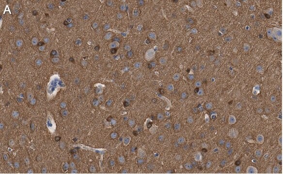
immunohistochemistry (formalin-fixed, paraffin-embedded sections): suitable
microarray: suitable
western blot: 1:1,000 using a fresh total bovine brain extract or an enriched microtubule protein preparation
동형
IgG1
UniProt 수납 번호
배송 상태
dry ice
저장 온도
−20°C
타겟 번역 후 변형
unmodified
유전자 정보
human ... MAPT(4137)
관련 카테고리
일반 설명
Monoclonal Anti- τ (TAU) (mouse IgG1 isotype) is derived from the hybridoma produced by the fusion of mouse myeloma cells and splenocytes from an immunized mouse. τ (TAU) proteins are a part of microtubule associated proteins (MAPs). They are densely found in neurons and in trace amounts in non-neuronal cells. In brain six isoforms of τ (TAU) proteins are present.
The antibody reacts exclusively with the chemically heterogeneous τ in both the phosphorylated and non-phosphorylated form. The antibody does not react with other MAPs or with tubulin. In immunohistochemical staining, it localizes τ along microtubules in axons, somata, dendrites and astrocytes, and on ribosomes. The antibody may be used for staining of τ in Alzheimer neurofibrillary tangles in sections of human brain tissue.
The best known microtubule associated proteins (MAPs) which copurify with microtubules are MAP2 and Tau. These two proteins are heat stable and stimulate formation of the microtubule polymer from purified tubulin subunits. Tau is chemically heterogenous, however, limited protolysis has demonstrated that the different eletrophoretic species are closely related. Tau is immunologically distinct from the other MAPs, namely MAP1, MAP2 and MAP5. Localization studies have demonstrated that Tau is intimately associated with the filamentous structures which compose the neurofibrillary tangles as found in an Alzheimer′s disease brain.
면역원
bovine microtubule-associated proteins (MAPs)
애플리케이션
Monoclonal Anti-τ (Tau) antibody has been used:
- in immunohistology
- in immunoblotting
- in dot blot
- in immunohistochemistry
Mouse monoclonal clone TAU-2 anti-Tau antibody maybe used to study microtubule associated proteins (MAP) expression and cytological localization in various tissue and cell lines, under different developmental and environmental circumstances.
생화학적/생리학적 작용
Monoclonal Anti- τ (TAU) is phosphatase independent; it will bind Tau proteins in either their phosphorylated or non-phosphorylated forms. It localizes Tau proteins along microtubules in axons, somata, dendrites, astrocytes and on ribosomes (polysomes). The best-known microtubule associated proteins (MAPs) which copurify with microtubules are MAP2 and Tau. These two proteins are heat stable and stimulate formation of the microtubule polymer from purified tubulin subunits. Tau is immunologically distinct from the other MAPs. Tau is intimately associated with the filamentous structures which compose the neurofibrillary tangles as found in an Alzheimer′s disease brain.
면책조항
Unless otherwise stated in our catalog or other company documentation accompanying the product(s), our products are intended for research use only and are not to be used for any other purpose, which includes but is not limited to, unauthorized commercial uses, in vitro diagnostic uses, ex vivo or in vivo therapeutic uses or any type of consumption or application to humans or animals.
적합한 제품을 찾을 수 없으신가요?
당사의 제품 선택기 도구.을(를) 시도해 보세요.
Storage Class Code
10 - Combustible liquids
WGK
WGK 3
Flash Point (°F)
Not applicable
Flash Point (°C)
Not applicable
가장 최신 버전 중 하나를 선택하세요:
시험 성적서(COA)
Lot/Batch Number
Chris R Guthrie et al.
Journal of molecular neuroscience : MN, 45(1), 32-41 (2011-02-23)
Lesions containing aggregated and hyperphosphorylated tau protein are characteristic of neurodegenerative tauopathies. We have developed a cellular model of pathological tau deposition and clearance by overexpressing wild type human tau in HEK293 cells. When proteasome activity is inhibited, HEK293/tau cells
R E Mrak et al.
Human pathology, 26(8), 816-823 (1995-08-01)
The roles of activated glia and of glial cytokines in the pathogenesis of Alzheimer's disease are reviewed. Interleukin-1 (IL-1), a microglia-derived acute phase cytokine, activates astrocytes and induces expression of the astrocyte-derived cytokine, S100 beta, which stimulates neurite growth (and
Cortical limb myoclonus in pathologically proven progressive supranuclear palsy.
Sharon Kemp et al.
Movement disorders : official journal of the Movement Disorder Society, 28(13), 1804-1806 (2013-10-15)
Severity of gliosis in Pick?s disease and frontotemporal lobar degeneration: tau-positive glia differentiate these disorders
Schofield E, et al.
Brain, 126(4), 827-840 (2003)
Melissa Broe et al.
Brain : a journal of neurology, 127(Pt 10), 2214-2220 (2004-07-30)
The main unifying feature of cases with frontotemporal dementia (FTD) is the pattern of brain atrophy. Surprisingly, there are a variety of underlying histopathologies in cases with the clinical features and typical pattern of atrophy characterizing FTD. This suggests that
활성 필터
자사의 과학자팀은 생명 과학, 재료 과학, 화학 합성, 크로마토그래피, 분석 및 기타 많은 영역을 포함한 모든 과학 분야에 경험이 있습니다..
고객지원팀으로 연락바랍니다.