
추천 제품


생물학적 소스
rabbit
Quality Level
결합
unconjugated
항체 형태
affinity isolated antibody
항체 생산 유형
primary antibodies
클론
polyclonal
양식
buffered aqueous solution
분자량
antigen 190 kDa
종 반응성
rat, chicken
향상된 검증
independent
Learn more about Antibody Enhanced Validation
농도
~0.5 mg/mL
기술
immunohistochemistry (formalin-fixed, paraffin-embedded sections): 1:200 using rat and chicken cerebellum sections
microarray: suitable
western blot: 1:500 using a rat brain extract
UniProt 수납 번호
배송 상태
dry ice
저장 온도
−20°C
타겟 번역 후 변형
unmodified
유전자 정보
human ... MYO5A(4644)
mouse ... Myo5a(17918)
rat ... Myo5a(25017)
일반 설명
Myosin Va (p190) is a member of the unconventional class of myosins, distinct from both the myosins I and myosin II. It is present in neuronal and non neuronal cells of the brain. Class V myosins are widely expressed actin-based motors. Class V myosins have two motor head domains typical of myosins, and an extended regulatory neck domain with six tandem IQ domains that bind multiple calmodulin light chains. In addition, myosin V contains a unique 400 amino acids globular tail domain that may direct myosin V to its target or determine the cargo to which it binds.
면역원
synthetic peptide located near the C-terminus of chicken myosin Va (amino acids 1705-1720 with N-terminally added lysine) conjugated to KLH. This sequence is identical in human, mouse and rat.
애플리케이션
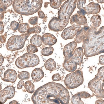
Anti-Myosin Va (LE-16) antibody produced in rabbit is used in immunoblotting and immunohistochemistry.
생화학적/생리학적 작용
Myosin Va is implicated in the regulation of vesicle trafficking in neurons and melanocytes. It regulates melanosome distribution along microfilaments. It is found in association with the centrosome at all stages of the cell cycle. In the interphase stage, myosin Va is found in pericentriolar material. During cell division, it is found in the cytoplasm and concentrates in a trail between migrating centrioles and in the mitotic spindle poles and spindle fibers.
물리적 형태
Solution in 0.01 M phosphate buffered saline, pH 7.4, containing 1% BSA and 15 mM sodium azide.
면책조항
Unless otherwise stated in our catalog or other company documentation accompanying the product(s), our products are intended for research use only and are not to be used for any other purpose, which includes but is not limited to, unauthorized commercial uses, in vitro diagnostic uses, ex vivo or in vivo therapeutic uses or any type of consumption or application to humans or animals.
적합한 제품을 찾을 수 없으신가요?
당사의 제품 선택기 도구.을(를) 시도해 보세요.
Storage Class Code
10 - Combustible liquids
WGK
WGK 3
Flash Point (°F)
Not applicable
Flash Point (°C)
Not applicable
A millennial myosin census
Berg JS, et al.
Molecular Biology of the Cell, 12(4), 780-794 (2001)
High affinity binding of brain myosin-Va to F-actin induced by calcium in the presence of ATP
Tauhata SBF, et al.
The Journal of Biological Chemistry, 276(43), 39812-39818 (2001)
Katharina N Richter et al.
Scientific reports, 8(1), 14838-14838 (2018-10-06)
Protein copy numbers can be measured by biochemical methods ranging from quantitative Western Blotting to several mass spectrometry approaches. Such methods only provide average copy numbers, obtained over large cell numbers. However, copy number estimates for single cells or single
Regulated conformation of myosin V
Wang F, et al.
The Journal of Biological Chemistry, 279(4), 2333-2336 (2004)
활성 필터
자사의 과학자팀은 생명 과학, 재료 과학, 화학 합성, 크로마토그래피, 분석 및 기타 많은 영역을 포함한 모든 과학 분야에 경험이 있습니다..
고객지원팀으로 연락바랍니다.