
추천 제품


생물학적 소스
rabbit
결합
alkaline phosphatase conjugate
항체 형태
affinity isolated antibody
항체 생산 유형
secondary antibodies
클론
polyclonal
양식
buffered aqueous glycerol solution
종 반응성
goat
기술
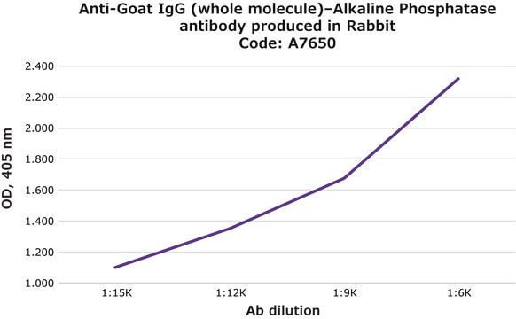
direct ELISA: 1:30,000
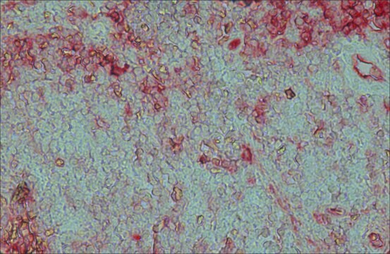
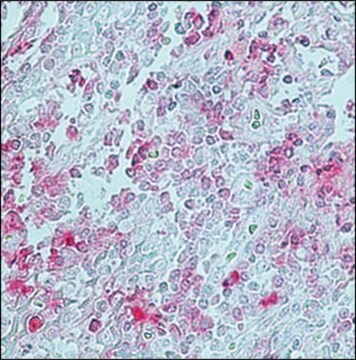
immunohistochemistry (formalin-fixed, paraffin-embedded sections): 1:50
western blot: 1:30,000
배송 상태
wet ice
저장 온도
2-8°C
타겟 번역 후 변형
unmodified
유사한 제품을 찾으십니까? 방문 제품 비교 안내
관련 카테고리
일반 설명
IgG antibodies regulate several functions such as complement activation and phagocytosis. Thus they play a crucial role in facilitating cytological immune responses. Polyclonal anti-goat IgG (whole molecule)–alkaline phosphatase antibody (diluted 1:15,000) can be used as a secondary antibody for immunoblotting of Sall4. This antibody can also be used in immunohistochemistry (diluted1:400) . Rabbit anti-goat IgG antibody reacts specifically with goat IgG and normal goat serum.
Primary goat antibodies are often used to study target proteins for various clinical and research purposes. Thus, secondary anti-goat antibody conjugated to a detectable substrate can be used to facilitate the accurate detection and localization of target proteins.
특이성
Binds all goat Igs
면역원
Purified goat IgG.
애플리케이션
Anti-Goat IgG (whole molecule)-Alkaline Phosphatase antibody is suitable for use in immunoblotting. The product can also be used for direct ELISA (1:30,000) and immunohistochemistry (1:50 using formalin-fixed, paraffin-embedded sections).
Detection of TLR2 and TLR4 in protein extracts from epidermal keratinocytes was performed by western blot using alkaline phosphatase conjugated rabbit anti-goat IgG as the secondary at a 1:2500 dilution.
물리적 형태
Solution in 0.05 M Tris buffer, pH 8.0, containing 1 mM MgCl2, 1% bovine serum albumin, 50% glycerol and 15 mM sodium azide.
면책조항
Unless otherwise stated in our catalog or other company documentation accompanying the product(s), our products are intended for research use only and are not to be used for any other purpose, which includes but is not limited to, unauthorized commercial uses, in vitro diagnostic uses, ex vivo or in vivo therapeutic uses or any type of consumption or application to humans or animals.
적합한 제품을 찾을 수 없으신가요?
당사의 제품 선택기 도구.을(를) 시도해 보세요.
Storage Class Code
10 - Combustible liquids
WGK
WGK 2
가장 최신 버전 중 하나를 선택하세요:
시험 성적서(COA)
Lot/Batch Number
이미 열람한 고객


Minoru Omi et al.
The Journal of comparative neurology, 519(13), 2615-2621 (2011-04-15)
The optic tectum is a visual center of nonmammalian vertebrates that receives retinal fibers in a retinotopic manner. It has been accepted that retinal fibers project to some superficial laminae of the tectum, but do not go through lamina g
Menhaj et al.
Planta, 209(4), 406-413 (1999-11-07)
An antibody was raised against the protein HL#2 which is a nuclear-encoded light-stress-induced protein of barley (Hordeum vulgare L.). The expression of the mRNA and the protein of HL#2 was determined under the influence of high light and methyl jasmonate.
Andor Pivarcsi et al.
International immunology, 15(6), 721-730 (2003-05-17)
Keratinocytes have the ability to kill pathogenic fungi and bacteria by producing antimicrobial substances. Recent studies suggest that microbial components use signaling molecules of the human Toll-like receptor (TLR) family to transduce signals in various cells. Here we provide evidence
L Moysset et al.
Planta, 213(4), 565-574 (2001-09-15)
The intracellular localization of phytochrome in the pulvini of Robinia pseudoacacia L. was analyzed by immunogold electron microscopy after red (R; 15 min) and far-red (FR; 5 min) irradiation 2 h after the beginning of the photoperiod. Screening of the
Tomasz Wasniewski et al.
Oncology reports, 34(5), 2760-2767 (2015-09-04)
In order to study lysophosphatidic acid (LPA) signaling associated with type 1 endometrial carcinoma (EC), we evaluated the LPA receptors (LPARs), autotaxin (ATX) and phospholipase A2 (PLA2) expression in EC and normal endometrium with correlation to clinicopathological features. We investigated LPAR1, LPAR2
활성 필터
자사의 과학자팀은 생명 과학, 재료 과학, 화학 합성, 크로마토그래피, 분석 및 기타 많은 영역을 포함한 모든 과학 분야에 경험이 있습니다..
고객지원팀으로 연락바랍니다.