
가격 및 재고 정보를 현재 이용할 수 없음 고객지원팀으로 연락바랍니다.
추천 제품


제품명
Anti-Procollagen Type I Antibody, CT, clone PCIDG10 (Ascites Free), clone PCIDG10, from mouse
생물학적 소스
mouse
Quality Level
항체 형태
purified immunoglobulin
항체 생산 유형
primary antibodies
클론
PCIDG10, monoclonal
종 반응성
mouse, human, rat, guinea pig
종 반응성(상동성에 의해 예측)
bovine (based on 100% sequence homology)
기술
ELISA: suitable
flow cytometry: suitable
immunocytochemistry: suitable
immunohistochemistry: suitable (paraffin)
동형
IgG1κ
NCBI 수납 번호
UniProt 수납 번호
배송 상태
dry ice
타겟 번역 후 변형
unmodified
유전자 정보
human ... COL1A1(1277)
관련 카테고리
일반 설명
Collagen alpha-1(I) chain (UniProt P02452; also known as Alpha-1 type I collagen) is encoded by the COL1A1 gene (Gene ID 1277) in human. Collagen is the major component of the extracellular matrix (ECM) and forms the fibrils of tendons, ligaments, and bones. Type I collagen consists of two alpha I chains and one alpha 2 chain. Alpha-1 type I collagen is initially produced as a 1464-amino acid prepro-form with a signal peptide sequence (a.a. 1-22) and two propeptide sequences (a.a. 23-161 and a.a. 1219 –1464), the removal of which yields the mature alpha-1(I) chain. The mature alpha-1(I) chain is composed mostly of a large triple-helical region (a.a. 179-1192) sandwiched between two nonhelical segments known as the N-terminal telopeptide (a.a. 162-178; numbering based on the prepro-form) and the C-terminal telopeptide (a.a. 1193-1218; numbering based on the prepro-form). Collagen can be extracted from tissue via either enzymatic or non-enzymatic means. Collagen extracted using the proteolytic enzyme pepsin corresponds to the large triple-helical region, referred to as atelocollagen because both the N- and C-terminal telopeptides have been cleaved off by pepsin. On the other hand, collagen preparations obtained with non-enzymatic means (e.g. by acid extraction) have the intact telopeptides at both ends.
특이성
Labels carboxy-terminal pro-peptide of collagen type I. Does not stain mature collagen fibers in tissue, but rather is localized intracellularly in cells producing pro-collagen I.
면역원
Epitope: C-terminal propeptide region
Human pro-collagen I.
애플리케이션
Immunohistochemistry Analysis: A 1:50 dilution from a representative lot detected Procollagen Type I in mouse skin, rat skin, and rat skeletal muscle tissue.
Immunocytochemistry Analysis: A representative lot detected type I procollagen immunoreactivity in human semitendinosus and gracilis tendon fibroblasts from patients undergoing reconstruction surgery after anterior cruciate ligament (ACL) rupture by fluorescent immunocytochemistry (Bayer, M.L., et al. (2012). Mech Ageing Dev. 133(5):246-254).
Flow Cytometry Analysis: A representative lot detected PICP+/CD45+ fibrocytes in lung cells from bleomycin-treated mice (Yeager, M.E., et al. (2012). Eur Respir J. 39(1):104-111).
Flow Cytometry Analysis: A representative lot detected higher numbers and percentages of circulating PICP+/CD45+ fibrocytes in peripheral blood samples from children/yound adults with pulmonary hypertension (PH) than in samples from healthy individuals (Reese, C., et al. (2014). Front Pharmacol. 5:141).
Immunohistochemistry Analysis: A representative lot detected cytoplasmic type I procollagen immunoreactivity in stromal cells from the lysed functionalis of frozen human menstrual endometria sections (Gaide Chevronnay, H.P., et al. (2009). Endocrinology. 150(11):5094-5105).
ELISA Analysis: A representative lot detected different age-dependency of procollagen type I C-terminal propeptide (PICP) immunoreactivity in the cruciate ligaments of osteoarthritis-/OA-prone Dunkin-Hartley (DH) guinea pigs and in age-matched Bristol strain 2 (BS2) control guinea pigs (Quasnichka, H.L., et al. (2005). Arthritis Rheum. 52(10):3100-3109).
Research Category
Cell Structure
Cell Structure
Research Sub Category
Adhesion (CAMs)
Adhesion (CAMs)
This Anti-Procollagen Type I Antibody, CT, clone PCIDG10 (Ascites Free) is validated for use in Immunohistochemistry (Paraffin), Immunocytochemistry, Flow Cytometry and ELISA for the detection of Procollagen Type I.
품질
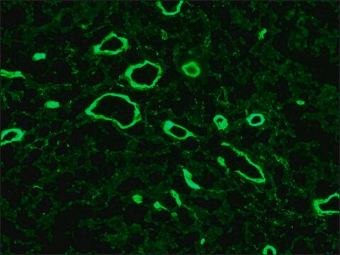
Evaluated by Immunohistochemistry in human bone tissue.
Immunohistochemistry Analysis: A 1:50 dilution of this antibody detected Procollagen Type I in human bone tissue.
Immunohistochemistry Analysis: A 1:50 dilution of this antibody detected Procollagen Type I in human bone tissue.
표적 설명
140 kDa calculated
물리적 형태
Format: Purified
Protein G Purified
Purified mouse monoclonal IgG1κ antibody in buffer containing 0.1 M Tris-Glycine (pH 7.4), 150 mM NaCl without preservatives.
저장 및 안정성
Stable for 1 year at -20°C from date of receipt.
Handling Recommendations: Upon receipt and prior to removing the cap, centrifuge the vial and gently mix the solution. Aliquot into microcentrifuge tubes and store at -20°C. Avoid repeated freeze/thaw cycles, which may damage IgG and affect product performance.
Handling Recommendations: Upon receipt and prior to removing the cap, centrifuge the vial and gently mix the solution. Aliquot into microcentrifuge tubes and store at -20°C. Avoid repeated freeze/thaw cycles, which may damage IgG and affect product performance.
기타 정보
Concentration: Please refer to lot specific datasheet.
면책조항
Unless otherwise stated in our catalog or other company documentation accompanying the product(s), our products are intended for research use only and are not to be used for any other purpose, which includes but is not limited to, unauthorized commercial uses, in vitro diagnostic uses, ex vivo or in vivo therapeutic uses or any type of consumption or application to humans or animals.
적합한 제품을 찾을 수 없으신가요?
당사의 제품 선택기 도구.을(를) 시도해 보세요.
Storage Class Code
12 - Non Combustible Liquids
WGK
WGK 1
Flash Point (°F)
Not applicable
Flash Point (°C)
Not applicable
시험 성적서(COA)
제품의 로트/배치 번호를 입력하여 시험 성적서(COA)을 검색하십시오. 로트 및 배치 번호는 제품 라벨에 있는 ‘로트’ 또는 ‘배치’라는 용어 뒤에서 찾을 수 있습니다.
J A McDonald et al.
The Journal of clinical investigation, 78(5), 1237-1244 (1986-11-01)
Excessive collagen deposition plays a critical role in the development of fibrosis, and early or active fibrosis may be more susceptible to therapeutic intervention than later stages of scarring. However, at present there is no simple method for assessing the
Héloïse P Gaide Chevronnay et al.
Endocrinology, 150(11), 5094-5105 (2009-10-13)
Coupling of focal degradation and renewal of the functional layer of menstrual endometrium is a key event of the female reproductive biology. The precise mechanisms by which the various endometrial cell populations control extracellular matrix (ECM) degradation in the functionalis
Yibo Yu et al.
International heart journal, 64(4), 632-640 (2023-07-31)
Atrial fibrillation (AF) is the most common arrhythmia that is harmful to human health. This study aims to explore the relationship between myosin light chain 4 (MYL4) and AF recurrence after radiofrequency ablation (RFA). Patients with AF (n = 85)
Monika L Bayer et al.
Mechanisms of ageing and development, 133(5), 246-254 (2012-03-08)
The aging process of tendon tissue is associated with decreased collagen content and increased risk for injuries. An essential factor in tendon physiology is transforming growth factor-β1 (TGF-β1), which is presumed to be reduced systemically with advanced age. The aim
Helen L Quasnichka et al.
Arthritis and rheumatism, 52(10), 3100-3109 (2005-10-04)
The influence of the cruciate ligaments in spontaneous osteoarthritis (OA) is not understood, although ligament rupture is known to cause secondary OA. Additionally, femoral notch narrowing at the anterior cruciate ligament (ACL) insertion site is associated with disease severity, but
활성 필터
자사의 과학자팀은 생명 과학, 재료 과학, 화학 합성, 크로마토그래피, 분석 및 기타 많은 영역을 포함한 모든 과학 분야에 경험이 있습니다..
고객지원팀으로 연락바랍니다.