추천 제품
생물학적 소스
mouse
Quality Level
결합
unconjugated
항체 형태
ascites fluid
항체 생산 유형
primary antibodies
클론
LH7.2, monoclonal
포함
15 mM sodium azide
종 반응성
human, goat, marmoset, monkey, guinea pig, sheep, pig, bovine
기술
dot blot: suitable
immunocytochemistry: suitable
indirect ELISA: suitable
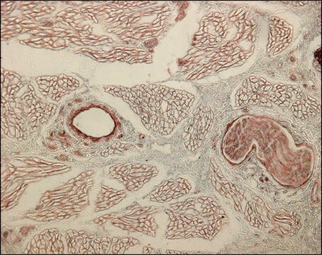
indirect immunofluorescence: 1:1,000 using human or other mammalian frozen sections
western blot: suitable
동형
IgG1
UniProt 수납 번호
배송 상태
dry ice
저장 온도
−20°C
타겟 번역 후 변형
unmodified
유전자 정보
human ... COL7A1(1294)
일반 설명
Collagen is a fibrous protein present in the extracellular framework of all vertebrates. Collagen type VII is the crucial component of anchoring fibrils and mutation in type VII collagen gene (COL7A1) leads to epidermolysis bullosa dystrophica. Monoclonal anti-collagen, type VII antibody can be used to distinguish invasive melanoma from non-invasive melanoma by visualizing the appearance and integrity of epidermal basement membrane. It can also be used in immunoblotting. Mouse anti-collagen type VII antibody reacts specifically with an epitope located on collagenase digested type VII collagen (150 kDa). The product also reacts with the basement membrane zone of stratified squamous epithelia of sheep, pig, bovine, guinea pig, goat and human.
Collagen type VII alpha 1 chain (COL7A1) is majorly expressed in skin, secreted by keratinocytes and fibroblasts. The gene is located on human chromosome 3p21.31.
면역원
insoluble fractions of neonatal human foreskin epidermal cells
애플리케이션
Monoclonal Anti-Collagen, Type VII antibody produced in mouse has been used in immuno-gold labelling.
Monoclonal anti-collagen type VII antibody can be used as a primary antibody in virus titration. It can also be used for the localization of type VII collagen in different immunochemical assays like dot blot, immunocytochemistry and ELISA.
생화학적/생리학적 작용
Collagen type VII alpha 1 chain (COL7A1) is required for epithelium-to-stroma anchorage in skin, mucosa and cornea. It is considered as an immediate-early response gene for transforming growth factor β (TGF-β)/SMAD (mothers against decapentaplegic) signaling pathway.
면책조항
Unless otherwise stated in our catalog or other company documentation accompanying the product(s), our products are intended for research use only and are not to be used for any other purpose, which includes but is not limited to, unauthorized commercial uses, in vitro diagnostic uses, ex vivo or in vivo therapeutic uses or any type of consumption or application to humans or animals.
Not finding the right product?
Try our 제품 선택기 도구.
Storage Class Code
12 - Non Combustible Liquids
WGK
nwg
Flash Point (°F)
Not applicable
Flash Point (°C)
Not applicable
시험 성적서(COA)
제품의 로트/배치 번호를 입력하여 시험 성적서(COA)을 검색하십시오. 로트 및 배치 번호는 제품 라벨에 있는 ‘로트’ 또는 ‘배치’라는 용어 뒤에서 찾을 수 있습니다.
이미 열람한 고객
Type VII Collagen in the Human Accommodation System: Expression in Ciliary Body, Zonules, and Lens Capsule
Wullink B, et al.
Investigative Ophthalmology & Visual Science, 59(2), 1075-1083 (2018)
Molecular basis of dystrophic epidermolysis bullosa: mutations in the type VII collagen gene (COL7A1).
Jarvikallio A, Pulkkinen L
Human Mutation, 10(5), 338-347 (1997)
Type VII collagen associated with the basement membrane of amniotic epithelium forms giant anchoring rivets which penetrate a massive lamina reticularis
Ockleford CD, et al.
Placenta, 34(9), 727-737 (2013)
COL7A1 editing via CRISPR/Cas9 in recessive dystrophic epidermolysis bullosa
Hainzl S, et al.
Molecular Therapy, 25(11), 2573-2584 (2017)
Daisuke Sawamura et al.
The Journal of investigative dermatology, 118(6), 967-971 (2002-06-13)
The expression of intradermally injected DNA by keratinocytes is found mainly in the upper and middle layers of the epidermis. To investigate the mechanism of this selective expression, we observed the sequential changes in the distribution of interleukin-6-expressing keratinocytes after
자사의 과학자팀은 생명 과학, 재료 과학, 화학 합성, 크로마토그래피, 분석 및 기타 많은 영역을 포함한 모든 과학 분야에 경험이 있습니다..
고객지원팀으로 연락바랍니다.


