일반 설명
The family of Early B-cell Factor (EBF) or Collier/Olf-1/EBF (COE) proteins, EBF1, EBF2, and EBF3, are ubiquitous zinc-binding transcription factors found in many species, including human, Drosophila melanogaster, and Caenorhabditis elegans. EBF1 is abundantly expressed in early B-cells, lymph node, spleen, and adipose tissue. Low levels of EBF1 have also been detected in the brain, heart, skeletal muscle, and kidney. EBF proteins are capable of forming homo- and heterodimers that bind to DNA at specific sites that include the sequence 5′-ATTCCCNNGGGAATT-3′. EBF1, the prototypical member of this family, may employ epigenetic mechanisms, such as DNA methylation and chromatin remodeling, to regulate genes involved in B-cell development such as Pax5. The second member of this family EBF2 is involved in bone development, adipogenesis, and CNS development, processes which may also hinge on EBF1. EBF3 has been characterized as a tumor suppressor protein, due to its role in regulating multiple genes such as those for cyclins and CDKs which control cell growth, differentiation, and apoptosis. However, emerging evidence indicates that all EBF proteins may play a role in the development of various cancers.
특이성
This antibody recognizes EBF-1 but not EBF-2 or EBF-3.
면역원
KLH-conjugated linear peptide corresponding to mouse EBF-1.
애플리케이션
Anti-EBF-1 Antibody is an antibody against EBF-1 for use in Western Blotting, IHC(P), Immunofluorescence, Dot Blot.
Research Category
Neuroscience
Research Sub Category
Developmental Neuroscience
Western Blot Analysis: 2 µg/mL from a representative lot detected EBF-1 in COS-7 cells transfected with EBF-3. (Image courtesy of Dr. Giacomo Consalez, San Raffaele Scientific Institute.)
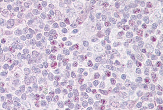
Immunohistochemistry Analysis: A 1:500 dilution from a representative lot detected EBF-1 in mouse cryosections of wild type spinal cord tissue. (Image courtesy of Dr. Giacomo Consalez, San Raffaele Scientific Institute.)
Immunofluorescence Analysis: A 1:500 dilution from a representative lot detected EBF-1 in COS-7 cells transfected with EBF expressing plasmids. (Image courtesy of Dr. Giacomo Consalez, San Raffaele Scientific Institute.)
Dot Blot Analysis: EBF-1, EBF-2, and EBF-3 peptides from a representative lot were probed with Anti-EBF-1 (1:100 dilution). No cross reactivity to peptides for EBF-3 & EBF-2 were observed.
품질
Evaluated by Western Blot in Raji cell lysate.
Western Blot Analysis: 2 µg/mL of this antibody detected EBF-1 in 10 µg of Raji cell lysate.
표적 설명
~66 kDa observed. Uncharacterized bands may appear between ~40 and ~170 kDa in some lysates.
물리적 형태
Affinity purified
Purified rabbit polyclonal in buffer containing 0.1 M Tris-Glycine (pH 7.4), 150 mM NaCl with 0.05% sodium azide.
저장 및 안정성
Stable for 1 year at 2-8°C from date of receipt.
분석 메모
Control
Raji cell lysate
기타 정보
Concentration: Please refer to the Certificate of Analysis for the lot-specific concentration.
면책조항
Unless otherwise stated in our catalog or other company documentation accompanying the product(s), our products are intended for research use only and are not to be used for any other purpose, which includes but is not limited to, unauthorized commercial uses, in vitro diagnostic uses, ex vivo or in vivo therapeutic uses or any type of consumption or application to humans or animals.


