詳細
Versican core protein (UniProt: Q9ERB4; also known as Chondroitin sulfate proteoglycan core protein 2, Glial hyaluronate-binding protein, GHAP, Large fibroblast proteoglycan, PG-M) is encoded by the Vcan (also known as Cspg2) gene (Gene ID: 114122) in rat. Versican is a large multi-domain chondroitin sulphate proteoglycan that is secreted by fibroblasts and other types of cells. Versican is present in most of the retina including the outer nuclear layer, the outer plexiform layer, and the inner plexiform layer. It is synthesized with a 20 amino acid signal peptide, which is subsequently cleaved. Veriscan plays a role in intercellular signaling and in connecting cells with the extracellular matrix and is also involved in axon regeneration, central nervous system and skeletal muscle development, and cell adhesion. It has unique N- and C-terminal globular regions, each with multiple motifs. It contains one C-type lectin domain, two EGF-like domains, one Ig-like V-type domain, two link domains and one Sushi (CCP/SCR) domains. It also has a N-terminal hyaluronic acid (HA) binding domain that has homology with the G1 and G2 domains of aggrecan. Four isoforms of versican termed as V0, V1, V2, and V3, have been reported with molecular weights of 370, 263, 180, and 74 kDa, respectively. Each isoform contains different lengths of the GAG binding region. V0 contains two GAG binding regions called CSalpha and CSbeta domains. V1 isoform has only CSbeta and V2 isoform has only CSalpha domain. V3 isoform contains only G1 and G3 domains and lacks GAG attachment sites. Defects in Vcan gene result in Wagner syndrome type 1, a type of vitreoretinopathy characterized by an optically empty vitreous cavity with fibrillary condensations and a pre-retinal avascular membrane.
免疫原
GST-tagged recombinant fragment corresponding to the 375 amino acids from the internal region of rat Versican.
アプリケーション
Anti-Versican, Beta Domain, Cat. No. ABT1370 is a rabbit polyclonal antibody that detects versican core protein and has been tested for use in Immunohistochemistry (Paraffin), Radioimmunoassay, and Western Blotting.
Radioimmunoassay Analysis: A representative lot detected Versican, Beta Domain in Postnatal rat brain (Milev, P., et. al. (1998). Biochem Biopys Res Commun. 247(2):207-12).
Immunohistochemistry Analysis: A representative lot detected Versican, Beta Domain in Embryonic day 16 rat eye, P0, P9 and adult rat retina (Popp, S., et. al. (2004). Exp Eye Res. 79(3):351-6) and Embryonic day 16, 19 rat brain and postnatal day 7 cerebellum, embryonic day 19 spinal cord (Popp, S., et. al. (2003). Dev Dyn. 227(1):143-9).
Western Blotting Analysis: A representative lot detected Versican, Beta Domain in purified proteoglycans from a PBS extract of 7-day rat brain with chondroitinase ABC treatment (Popp, S., et. al. (2003). Dev Dyn. 227(1):143-9).
品質
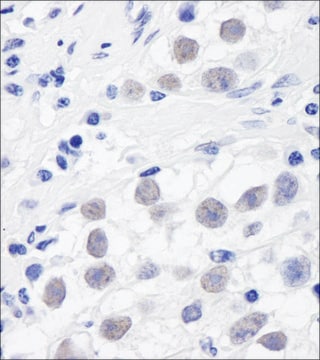
Evaluated by Immunohistochemistry in rat eye retina tissue.
Immunohistochemistry Analysis: A 1:1,000 dilution of this antibody detected Versican, Beta Domain in rat eye retina tissue.
ターゲットの説明
300 kDa calculated.
その他情報
Concentration: Please refer to lot specific datasheet.


