MABC87
Anti-CD148/DEP-1 Antibody, clone Ab1 (Azide Free)
clone Ab 1, from mouse
Sinonimo/i:
CD148, DEP-1, Receptor-type tyrosine-protein phosphatase eta, Protein-tyrosine phosphatase eta, R-PTP-eta, Density-enhanced phosphatase 1, DEP1, HPTP eta, Protein-tyrosine phosphatase receptor type J, R-PTP-J
About This Item
Prodotti consigliati
Origine biologica
mouse
Livello qualitativo
Forma dell’anticorpo
purified immunoglobulin
Tipo di anticorpo
primary antibodies
Clone
Ab 1, monoclonal
Reattività contro le specie
human, mouse
tecniche
immunohistochemistry: suitable (paraffin)
immunoprecipitation (IP): suitable
inhibition assay: suitable
western blot: suitable
Isotipo
IgG1κ
N° accesso NCBI
N° accesso UniProt
Condizioni di spedizione
wet ice
modifica post-traduzionali bersaglio
unmodified
Informazioni sul gene
human ... PTPRJ(5795)
Descrizione generale
Immunogeno
Applicazioni
Apoptosis & Cancer
Apoptosis - Additional
Western Blot Analysis: A representative lot of this antibody detected CD148/DEP-1 in Mouse kidney lysates (Takahashi, T., et al., (2006) Blood. 108:1234-1242).
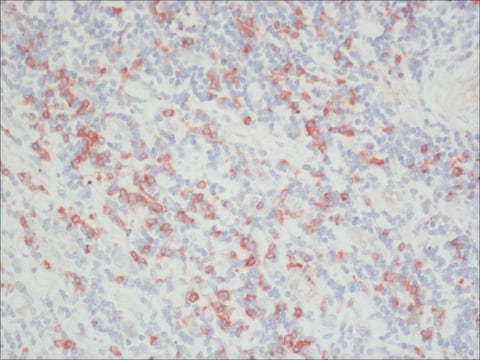
Immunohistochemistry Analysis: A 1:50 dilution of a representative lot detected CD148/DEP-1 in Epithelial Cells of human prostate tissue.
Immunohistochemistry Analysis: A representative lot of this antibody detected CD148/DEP-1 in frozen mouse embryos. (Takahashi, T., et al., (2003) MOLECULAR AND CELLULAR BIOLOGY. 23(5):1817–1831).
Immunohistochemistry Analysis: A representative lot of this antibody detected CD148/DEP-1 in various mouse tissues such as kidney, cortex and cornea. (Takahashi, T., et al., (2006) Blood. 108:1234-1242).
Inhibition Assay: A representative lot of this antibody inhibited proliferation of HRMEC′s (Primary human renal microvascular endothelial cells) (Takahashi, T., et al., (2006) Blood. 108:1234-1242).
Immunoprecipitation Analysis: A representative lot of this antibody immunoprecipitated CD148/DEP-1 from Huvec and RMEC cell lysates (Takahashi, T., et al., (1999) J. Am. Soc. Nephrol. 10: 2135–2145).
Qualità
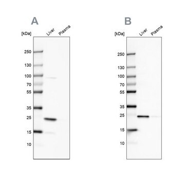
Western Blotting Analysis: 0.5 µg/mL of this antibody detected CD148/DEP-1 in 10 µg of HeLa cell lysate.
Descrizione del bersaglio
This protein is highly glycosylated and may run higher than the calculated molecular weight of 146 kDa (isoform 1) or 57 kDa (isoform 2). Uncharacterized bands may be observed in some lysates.
Stato fisico
Stoccaggio e stabilità
Handling Recommendations: Upon receipt and prior to removing the cap, centrifuge the vial and gently mix the solution. Aliquot into microcentrifuge tubes and store at -20°C. Avoid repeated freeze/thaw cycles, which may damage IgG and affect product performance.
Altre note
Esclusione di responsabilità
Not finding the right product?
Try our Motore di ricerca dei prodotti.
Codice della classe di stoccaggio
12 - Non Combustible Liquids
Classe di pericolosità dell'acqua (WGK)
WGK 2
Punto d’infiammabilità (°F)
Not applicable
Punto d’infiammabilità (°C)
Not applicable
Certificati d'analisi (COA)
Cerca il Certificati d'analisi (COA) digitando il numero di lotto/batch corrispondente. I numeri di lotto o di batch sono stampati sull'etichetta dei prodotti dopo la parola ‘Lotto’ o ‘Batch’.
Possiedi già questo prodotto?
I documenti relativi ai prodotti acquistati recentemente sono disponibili nell’Archivio dei documenti.
Il team dei nostri ricercatori vanta grande esperienza in tutte le aree della ricerca quali Life Science, scienza dei materiali, sintesi chimica, cromatografia, discipline analitiche, ecc..
Contatta l'Assistenza Tecnica.


