Passa al contenuto
Merck
Tutte le immagini(4)

Documenti

MAB4190

Sigma-Aldrich

Anti-Ki-67 Antibody, clone Ki-S5

clone Ki-S5, Chemicon®, from mouse

Autenticatiper visualizzare i prezzi riservati alla tua organizzazione & contrattuali


About This Item

Codice UNSPSC:
12352203
eCl@ss:
32160702
NACRES:
NA.41

Origine biologica

mouse

Livello qualitativo

Forma dell’anticorpo

purified immunoglobulin

Tipo di anticorpo

primary antibodies

Clone

Ki-S5, monoclonal

Reattività contro le specie

human

Confezionamento

antibody small pack of 25 μg

Produttore/marchio commerciale

Chemicon®

tecniche

flow cytometry: suitable
immunocytochemistry: suitable
immunohistochemistry (formalin-fixed, paraffin-embedded sections): suitable
western blot: suitable

Isotipo

IgG1

N° accesso NCBI

N° accesso UniProt

Condizioni di spedizione

ambient

Temperatura di conservazione

2-8°C

modifica post-traduzionali bersaglio

unmodified

Informazioni sul gene

human ... MKI67(4288)

Descrizione generale

Ki67 antigen is the prototypic cell cycle related nuclear protein, expressed by proliferating cells in all phases of the active cell cycle (G1, S, G2 and M phase). It is absent in resting (G0) cells. Ki67 antibodies are useful in establishing the cell growing fraction in neoplasms (immunohistochemically quantified by determining the number of Ki67 positive cells among the total number of resting cells = Ki67 index). In neoplastic tissues the prognostic value is comparable to the tritiated thymidine labelling index. The correlation between low Ki67 index and histologically low grade tumours is strong. Ki67 is routinely used as a marker of cell cycling and proliferation. This specificity of Ki-S5 antibody for proliferating cells might make it a useful tool for determination of the proliferative fraction in tumors such as Non-Hodgkin′s lymphoma (Kreipe et al., 1993), mammary carcinomas (Mauri et al., 1984), ovarian tumors (Ballin et al., 1994) and prostate cancer.

Specificità

In Immunoprecipitation and Western blot experiments the Ki-S5 antibody recognizes a cell-cycle-associated protein of 345 kD and 395 kD (Kreipe et al., 1993) identical with the Ki-67 antigen. The antibody binds to a formalin-resistant epitope of the Ki-67 antigen. The immunoreactivity of Ki-S5 is confined to the nuclei of proliferating cells and no crossreactivity with cytoplasmic antigens of epithelial occurs even after antigen retrieval (Mauri et al., 1984). A comparison of immunohistochemical labeling of fresh and fixed tissue samples of NHL showed that identical results were obtained with Ki-67 and Ki-S5 (Kreipe et al., 1993). The Ki-67 antigen is preferentially expressed during late G1, S, G2 and M phase of the cell cycle, while resting, non-cycling cells (G0 phase) lack the Ki-67 antigen. In addition, constantly proliferating cells (e.g. cell lines) react positively to Ki-S5 during the entire cell-cycle. This specificity of Ki-S5 antibody for proliferating cells might make it a useful tool for determination of the proliferative fraction in tumors such as Non-Hodgkin′s lymphoma (Kreipe et al., 1993), mammary carcinomas (Mauri et al., 1984), ovarian tumors (Ballin et al., 1994) and prostate cancer.

Immunogeno

Nuclear protein preparation from the human cell line U937.50.

Applicazioni

Anti-Ki-67 Antibody, clone Ki-S5 is a high quality Mouse Monoclonal Antibody for the detection of Ki-67 & has been validated in FC, ICC, IHC, IHC(P) & WB.
Immunocytochemistry:
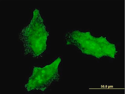
A 5-10 μg/mL concentration of a previous lot was used in IC.

Immunohistochemistry:
A 5-10 μg/mL concentration of a previous lot was used in IH.

Immunohistochemistry:
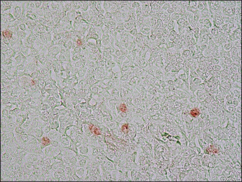
5-10 µg/mL on formalin fixed paraffin tissue. Citrate buffer-microwave antigen retrieval

Flow Cytometry:
A previous lot of this antibody was used in FC.

Western blot:
1-10 µg/mL

Optimal working dilutions must be determined by end user.
Research Category
Epigenetics & Nuclear Function
Research Sub Category
Cell Cycle, DNA Replication & Repair

Qualità

Evaluated by Western Blot on A431 lysates.

Western Blot Analysis:
1:500 dilution of this antibody detected cell-cycle-associated protein of 345 kDa and 395 kDa on 10 μg of A431 lysates.

Descrizione del bersaglio

345 & 395 kDa

Stato fisico

Format: Purified
Mouse monoclonal IgG1 in buffer containing 0.02 M Phosphate buffer, 0.25 M NaCl, pH 7.6 with 0.1% sodium azide.
Protein A purified

Stoccaggio e stabilità

Stable for 1 year at 2-8ºC from date of receipt.

Risultati analitici

Control
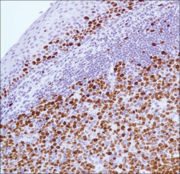
Tonsil tissue, A431 cell lysate

Altre note

Concentration: Please refer to the Certificate of Analysis for the lot-specific concentration.

Note legali

CHEMICON is a registered trademark of Merck KGaA, Darmstadt, Germany

Esclusione di responsabilità

Unless otherwise stated in our catalog or other company documentation accompanying the product(s), our products are intended for research use only and are not to be used for any other purpose, which includes but is not limited to, unauthorized commercial uses, in vitro diagnostic uses, ex vivo or in vivo therapeutic uses or any type of consumption or application to humans or animals.

Not finding the right product?  

Try our Motore di ricerca dei prodotti.

Raccomandato

N° Catalogo
Descrizione
Determinazione del prezzo

Codice della classe di stoccaggio

10 - Combustible liquids

Classe di pericolosità dell'acqua (WGK)

WGK 2

Punto d’infiammabilità (°F)

Not applicable

Punto d’infiammabilità (°C)

Not applicable


Certificati d'analisi (COA)

Cerca il Certificati d'analisi (COA) digitando il numero di lotto/batch corrispondente. I numeri di lotto o di batch sono stampati sull'etichetta dei prodotti dopo la parola ‘Lotto’ o ‘Batch’.

Possiedi già questo prodotto?

I documenti relativi ai prodotti acquistati recentemente sono disponibili nell’Archivio dei documenti.

Visita l’Archivio dei documenti

Paula Rocktäschel et al.
Epilepsy & behavior : E&B, 101(Pt B), 106581-106581 (2019-11-26)
Tuberous sclerosis complex (TSC) is a neurodevelopmental disorder caused by deletions in the TSC1 or TSC2 genes that is associated with epilepsy in up to 90% of patients. Seizures are suggested to start in benign brain tumors, cortical tubers, or
S Blondel et al.
Cell death & disease, 7, e2105-e2105 (2016-02-20)
Hutchinson-Gilford progeria syndrome (HGPS) is a rare genetic disorder characterized by a dramatic appearance of premature aging. HGPS is due to a single-base substitution in exon 11 of the LMNA gene (c.1824C>T) leading to the production of a toxic form
Sandra Haider et al.
Endocrinology, 155(1), 263-274 (2013-11-06)
Failures in human extravillous trophoblast (EVT) development could be involved in the pathogenesis of pregnancy diseases. However, the underlying mechanisms have been poorly characterized. Here, we provide evidence that Notch signaling could represent a key regulatory pathway controlling trophoblast proliferation
Identification of a Src tyrosine kinase/SIAH2 E3 ubiquitin ligase pathway that regulates C/EBP? expression and contributes to transformation of breast tumor cells.
Sarkar, TR; Sharan, S; Wang, J; Pawar, SA; Cantwell, CA; Johnson, PF; Morrison, DK; Wang et al.
Molecular and cellular biology null
Saskia Grudzenski et al.
Stem cell research & therapy, 8(1), 96-96 (2017-04-28)
In the field of experimental stem cell therapy, intra-arterial (IA) delivery yields the best results concerning, for example, migrated cell number at the targeted site. However, IA application also appears to be associated with increased mortality rates and infarction. Since

Il team dei nostri ricercatori vanta grande esperienza in tutte le aree della ricerca quali Life Science, scienza dei materiali, sintesi chimica, cromatografia, discipline analitiche, ecc..

Contatta l'Assistenza Tecnica.