


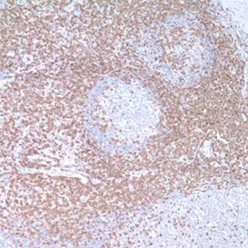


Based on the information available, this version does recognize the epsilon subunit. This conclusion is drawn from the fact that the CD3 epsilon, also known as the cytoplasmic CD3 antibody, exhibits a cytoplasmic staining pattern with perinuclear Golgi accentuation and very rarely membranous. Typically, this antibody is utilized for identifying T cell and NK cell lymphomas in FFPE tissues (please refer to representative staining photos). While the Cd3 surface antibody is commonly used in flow cytometry for membranous CD3 detection, the CD3 epsilon cytoplasmic is generally used in FFPE tissue, as is often done for IHC.
Select a Size
7 ML
HUF 135,000.00
1 ML
HUF 267,000.00
All Photos(1)
Select a Size
Change View
7 ML
HUF 135,000.00
1 ML
HUF 267,000.00
About This Item
UNSPSC Code:
12352203
NACRES:
NA.41
Recommended Products
biological source
rabbit
Quality Level
100
500
conjugate
unconjugated
antibody form
culture supernatant
antibody product type
primary antibodies
clone
MRQ-39, monoclonal
description
For In Vitro Diagnostic Use in Select Regions (See Chart)
form
buffered aqueous solution
species reactivity
human
packaging
pkg of 0.1 mL concentrate (103r-94)
pkg of 0.5 mL concentrate (103R-95)
pkg of 1.0 mL concentrate (103R-96)
pkg of 1.0 mL predilute (103R-97)
pkg of 7.0 mL predilute (103R-98)
manufacturer/tradename
Cell Marque®
IVD
for in vitro diagnostic use
technique(s)
immunohistochemistry (formalin-fixed, paraffin-embedded sections): 1:100-1:500 (concentrated)
isotype
IgG1
control
tonsil
shipped in
wet ice
storage temp.
2-8°C
visualization
membranous
Gene Information
human ... CD3E(916)
Related Categories
General description
Anti-CD3 has been considered the best all around T-cell marker. This antibody reacts with an antigen present in early thymocytes. The positive staining of this marker may represent a sign of early commitment to the T-Cell lineage.
Quality
 IVD |  IVD |  IVD |  RUO |
Linkage
CD3 Positive Control Slides, Product No. 103S, are available for immunohistochemistry (formalin-fixed, paraffin-embedded sections).
Physical form
Solution in Tris Buffer, pH 7.3-7.7, with 1% BSA and <0.1% Sodium Azide
Preparation Note
Download the IFU specific to your product lot and formatNote: This requires a keycode which can be found on your packaging or product label.
Other Notes
For Technical Service please contact: 800-665-7284 or email: [email protected]
Legal Information
Cell Marque is a registered trademark of Merck KGaA, Darmstadt, Germany
Not finding the right product?
Try our Product Selector Tool.
Choose from one of the most recent versions:
Certificates of Analysis (COA)
Lot/Batch Number
Don't see the Right Version?
If you require a particular version, you can look up a specific certificate by the Lot or Batch number.
Already Own This Product?
Find documentation for the products that you have recently purchased in the Document Library.
SM Denning, et al.
Leucocyte Typing III, 144-147 (1987)
Kennosuke Karube et al.
The American journal of surgical pathology, 27(10), 1366-1374 (2003-09-26)
We studied the morphologic, immunohistochemical, and clinical characteristics of 158 cases of lymphoblastic lymphoma. Based on immunophenotyping and cell lineage, cases were classified into B-cell type (CD20,CD19 or CD79a+, n = 53), T-cell type (surface CD3+, n = 84), and
Ulla Axdorph et al.
APMIS : acta pathologica, microbiologica, et immunologica Scandinavica, 110(5), 379-390 (2002-06-22)
Morphologically, T-cell-rich B-cell lymphoma (TCRB-NHL) may be indistinguishable from Hodgkin's disease (HD). Immunophenotyping may be helpful in the separation of these entities. TCRB-NHL is occasionally misdiagnosed and treated as HD. However, information is limited regarding clinical characteristics and outcome of
Ahmet Dogan et al.
The American journal of surgical pathology, 27(7), 903-911 (2003-06-27)
Occasionally, primary large B-cell lymphomas (LBLs) arising in the spleen present with a micronodular pattern involving the splenic white pulp but sparing the red pulp. Histologically, the nodules contain scattered large B cells in a background of numerous T cells
E A Clark et al.
Immunology today, 10(7), 225-228 (1989-07-01)
During 1987, striking advances were made in defining the receptors and ligands for cell-to-cell adhesion interactions involving leukocytes. In 1988, two major leukocyte differentiation antigens, CD10 (cALLA) and CD45 (LCA, T200), were shown to be enzymes while two other markers
-
Could you confirm if the CD3 clone (Catalog #103R-96) recognizes epsilon?
1 answer-
Helpful?
-
Active Filters
Our team of scientists has experience in all areas of research including Life Science, Material Science, Chemical Synthesis, Chromatography, Analytical and many others.
Contact Technical Service