MABE286
Anti-Replication Protein A Antibody, clone RPA34-19
clone RPA34-19, from mouse
别名:
Replication protein A 32 kDa subunit, RP-A p32, Replication factor A protein 2, RF-A protein 2, Replication protein A 34 kDa subunit, RP-A p34
登录查看公司和协议定价
所有图片(3)
About This Item
分類程式碼代碼:
12352203
eCl@ss:
32160702
NACRES:
NA.41
推荐产品
生物源
mouse
品質等級
抗體表格
purified antibody
抗體產品種類
primary antibodies
無性繁殖
RPA34-19, monoclonal
物種活性
human
技術
immunocytochemistry: suitable
immunohistochemistry: suitable
western blot: suitable
同型
IgG1κ
NCBI登錄號
UniProt登錄號
運輸包裝
wet ice
目標翻譯後修改
unmodified
基因資訊
human ... RPA2(6118)
一般說明
RPA (RP-A p32) is a heterotrimeric protein complex that binds specifically to single-stranded DNA (ssDNA). It is composed of three subunits: RPA1 (70 kDa), RPA2 (32 kDa), and RPA3 (14 kDa). RPA plays multiple roles in DNA replication. At the onset of DNA replication, RPA is loaded onto chromatin, and is needed for subsequent loading of DNA polymerase and other replication proteins to initiate DNA replication. After replication begins, RPA moves with replication forks, stabilizing ssDNA and assisting in DNA synthesis. In addition to its replication function, RPA is also known to play essential roles in damage repair and recombination. The 32 kDa subunit is phosphorylated by the cdc2 family of kinases when cells enter S-phase; and by ATM, ATR, and DNA-PK proteins in response to DNA damage.
免疫原
Replication Protein A purified from U293 cells.
應用
Anti-Replication Protein A Antibody, clone RPA34-19 is a Mouse Monoclonal Antibody for detection of Replication Protein A also known as Replication protein A 32 kDa subunit, RP-A p32 & has been validated in WB, ICC & IHC.
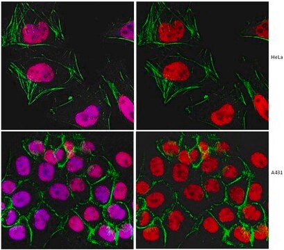
Immunocytochemistry Analysis: A 1:250 dilution from a reprsentative lot detected Replication Protein A in A431 cells.
Immunohistochemistry Analysis: A 1:5 dilution from a representative lot detected Replication Protein A in human placental chorionic villi and in human colorectal adenocarcinoma tissue.
Immunohistochemistry Analysis: A 1:5 dilution from a representative lot detected Replication Protein A in human placental chorionic villi and in human colorectal adenocarcinoma tissue.
Research Category
Epigenetics & Nuclear Function
Epigenetics & Nuclear Function
Research Sub Category
Cell Cycle, DNA Replication & Repair
Cell Cycle, DNA Replication & Repair
品質
Evaluated by Western Blot in HeLa cell lysate.
Western Blot Analysis: A 1:2,000 dilution of this antibody detected Replication Protein A in 10 µg of HeLa cell lysate.
Western Blot Analysis: A 1:2,000 dilution of this antibody detected Replication Protein A in 10 µg of HeLa cell lysate.
標靶描述
~34 kDa observed. This protein has 3 isoforms: Isoform 1 (~29 kDa), Isoform 2 (~30 kDa), and Isoform 3 (~39 kDa).
外觀
Protein G Purified
Format: Purified
Purified mouse monoclonal IgG1κ in buffer containing 0.1 M Tris-Glycine (pH 7.4), 150 mM NaCl with 0.05% sodium azide.
儲存和穩定性
Stable for 1 year at 2-8°C from date of receipt.
分析報告
Control
HeLa cell lysate
HeLa cell lysate
免責聲明
Unless otherwise stated in our catalog or other company documentation accompanying the product(s), our products are intended for research use only and are not to be used for any other purpose, which includes but is not limited to, unauthorized commercial uses, in vitro diagnostic uses, ex vivo or in vivo therapeutic uses or any type of consumption or application to humans or animals.
Not finding the right product?
Try our 产品选型工具.
儲存類別代碼
12 - Non Combustible Liquids
水污染物質分類(WGK)
WGK 1
閃點(°F)
Not applicable
閃點(°C)
Not applicable
Motohiro Yamauchi et al.
DNA repair, 7(3), 405-417 (2008-02-06)
Several DNA damage checkpoint factors form nuclear foci in response to ionizing radiation (IR). Although the number of the initial foci decreases concomitantly with DNA double-strand break repair, some fraction of foci persists. To date, the physiological role of the
Janna Luessing et al.
iScience, 25(7), 104536-104536 (2022-06-28)
Abscission, the final stage of cytokinesis, occurs when the cytoplasmic canal connecting two emerging daughter cells is severed either side of a large proteinaceous structure, the midbody. Here, we expand the functions of ATR to include a cell-cycle-specific role in
我们的科学家团队拥有各种研究领域经验,包括生命科学、材料科学、化学合成、色谱、分析及许多其他领域.
联系技术服务部门


