
推荐产品


生物来源
mouse
质量水平
偶联物
unconjugated
抗体形式
purified antibody
抗体产品类型
primary antibodies
克隆
9D9F9, monoclonal
分子量
calculated mol wt 152.76 kDa
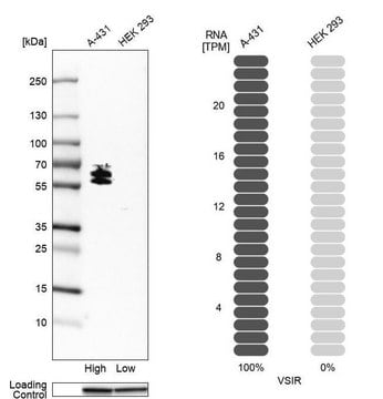
observed mol wt ~190 kDa
纯化方式
using protein G
种属反应性
human
包装
antibody small pack of 100 μL
技术
immunocytochemistry: suitable
immunohistochemistry (formalin-fixed, paraffin-embedded sections): suitable
immunoprecipitation (IP): suitable
western blot: suitable
同位素/亚型
IgG1
表位序列
Extracellular domain
Protein ID登记号
UniProt登记号
运输
2-8°C
靶向翻译后修饰
unmodified
基因信息
human ... FLT4(2324)
一般描述
Vascular endothelial growth factor receptor 3 (UniProt: P35916; also known as EC:2.7.10.1, VEGFR-3, Fms-like tyrosine kinase 4, FLT-4, Tyrosine-protein kinase receptor FLT4) is encoded by the FLT4 (also known as VEGFR3) gene (Gene ID: 2324) in human. FLT-4 is a single-pass type I membrane glycoprotein that is synthesized with a signal peptide (aa 1-24), which is subsequently cleaved off to generate the mature form that contains an extracellular domain (aa 25-775), a transmembrane domain (aa 776-796), and cytoplasmic domain (aa 797-1363). It is expressed in endothelial cells and is also detected in fetal spleen, lung, and brain. In adults it is detected in liver, muscle, thymus, placenta, lung, testis, ovary, prostate, heart, and kidney. FLT-4 has tyrosine-protein kinase activity and acts as a receptor for VEGFC and VEGFD. It plays a role in adult lymphangiogenesis and in the development of the vascular network and the cardiovascular system during embryonic development. Its secreted form functions as a decoy receptor for VEGFC and/or VEGFD and play an important role as a negative regulator of VEGFC-mediated lymphangiogenesis and angiogenesis. Its major function is to promote proliferation, survival, and migration of endothelial cells, and regulates angiogenic sprouting. FLT-4 undergoes trans auto-phosphorylation on tyrosine residues upon ligand binding. Phosphorylation at tyrosine 1068 is required for autophosphorylation at additional tyrosine residues. Phosphorylation at tyrosine 1063 and 1337 are important for interaction with Crk adaptor proteins and subsequent activation of MAPK8. In addition, its phosphorylation at tyrosine 1230, 1231, and 1337 are reported to be important for interaction with Grb2 and subsequent activation of the AKT1 and ERK1/ERK2 signaling pathways.
特异性
Clone 9D9F9 is a mouse monoclonal antibody that detects Vascular endothelial growth factor receptor 3 (VEGFR-3). It targets an epitope within the extracellular domain.
免疫原
A recombinant fragment corresponding to the extracellular domain of human Vascular endothelial growth factor receptor 3 (VEGFR-3).
应用
Quality Control Testing
Evaluated by Western Blotting in lysate from HEK293 cells over-expressing VEGFR-3.
Western Blotting Analysis (WB): A 1:500 dilution of this antibody detected Vascular endothelial growth factor receptor 3 (VEGFR-3) in lysate from HEK293 cells over-expressing human VEGFR-3.
Tested Applications
Immunoprecipitation Analysis: A representative lot immunoprecipitated VEGF Receptor-3 in Immunoprecipitation applications (Bando, H., et al. (2004). Int J Cancer; 111(2):184-91).
ELISA Analysis: A representative lot detected VEGF Receptor-3 in ELISA applications (Bando, H., et al. (2004). Int J Cancer; 111(2):184-91).
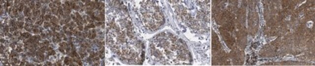
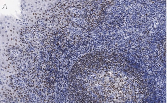
Immunohistochemistry (Paraffin) Analysis: A 1:250 dilution from a representative lot detected VEGF Receptor-3 in human placenta and human kidney tissue sections.
Western Blotting Analysis: A representative lot detected VEGF Receptor-3 in Western Blotting applications (Bando, H., et al. (2004). Int J Cancer; 111(2):184-91; Zhang, Y., et al. (2018). Nat Commun.; 9(1):1296).
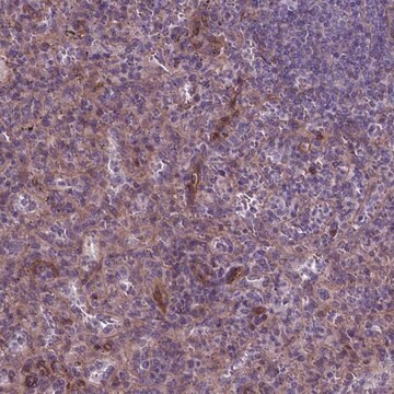
Immunohistochemistry Applications: A representative lot detected VEGF Receptor-3 in Immunohistochemistry applications (Bando, H., et al. (2004). Int J Cancer; 111(2):184-91; Jussila, L. et al. (1998). Cancer Res.; 58(8):1599-604; Su, C., et al. (2011). J Exp Clin Cancer Res.; 30(1):85; Valtola, R., et al. (1999). Am J. Pathol.; 154(5):1381-90).
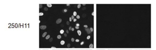
Immunocytochemistry Analysis: A 1:100 dilution from a representative lot detected VEGF Receptor-3 in human umbilical vascular endothelial cells (HUVEC).
Note: Actual optimal working dilutions must be determined by end user as specimens, and experimental conditions may vary with the end user
Evaluated by Western Blotting in lysate from HEK293 cells over-expressing VEGFR-3.
Western Blotting Analysis (WB): A 1:500 dilution of this antibody detected Vascular endothelial growth factor receptor 3 (VEGFR-3) in lysate from HEK293 cells over-expressing human VEGFR-3.
Tested Applications
Immunoprecipitation Analysis: A representative lot immunoprecipitated VEGF Receptor-3 in Immunoprecipitation applications (Bando, H., et al. (2004). Int J Cancer; 111(2):184-91).
ELISA Analysis: A representative lot detected VEGF Receptor-3 in ELISA applications (Bando, H., et al. (2004). Int J Cancer; 111(2):184-91).
Immunohistochemistry (Paraffin) Analysis: A 1:250 dilution from a representative lot detected VEGF Receptor-3 in human placenta and human kidney tissue sections.
Western Blotting Analysis: A representative lot detected VEGF Receptor-3 in Western Blotting applications (Bando, H., et al. (2004). Int J Cancer; 111(2):184-91; Zhang, Y., et al. (2018). Nat Commun.; 9(1):1296).
Immunohistochemistry Applications: A representative lot detected VEGF Receptor-3 in Immunohistochemistry applications (Bando, H., et al. (2004). Int J Cancer; 111(2):184-91; Jussila, L. et al. (1998). Cancer Res.; 58(8):1599-604; Su, C., et al. (2011). J Exp Clin Cancer Res.; 30(1):85; Valtola, R., et al. (1999). Am J. Pathol.; 154(5):1381-90).
Immunocytochemistry Analysis: A 1:100 dilution from a representative lot detected VEGF Receptor-3 in human umbilical vascular endothelial cells (HUVEC).
Note: Actual optimal working dilutions must be determined by end user as specimens, and experimental conditions may vary with the end user
Anti-VEGF Receptor-3, clone 9D9F9, Cat. No. MAB3757-I, is a mouse monoclonal antibody that detects VEGF receptor 3 and is tested for use in ELISA, Immunocytochemistry, Immunohistochemistry (Paraffin), Immunoprecipitation, and Western Blotting.
外形
Purified mouse monoclonal antibody IgG1 in buffer containing 0.1 M Tris-Glycine (pH 7.4), 150 mM NaCl with 0.05% sodium azide.
储存及稳定性
Recommend storage at +2°C to +8°C. For long term storage antibodies can be kept at -20°C. Avoid repeated freeze-thaws.
其他说明
Concentration: Please refer to the Certificate of Analysis for the lot-specific concentration.
免责声明
Unless otherwise stated in our catalog or other company documentation accompanying the product(s), our products are intended for research use only and are not to be used for any other purpose, which includes but is not limited to, unauthorized commercial uses, in vitro diagnostic uses, ex vivo or in vivo therapeutic uses or any type of consumption or application to humans or animals.
未找到合适的产品?
试试我们的产品选型工具.
储存分类代码
12 - Non Combustible Liquids
WGK
WGK 1
闪点(°F)
Not applicable
闪点(°C)
Not applicable
我们的科学家团队拥有各种研究领域经验,包括生命科学、材料科学、化学合成、色谱、分析及许多其他领域.
联系客户支持