Přejít k obsahu
Merck
Všechny fotografie(1)

Key Documents

S193

Sigma-Aldrich

Anti-Synapsin I antibody produced in rabbit

affinity isolated antibody, lyophilized powder

Synonyma:

Anti-EPILX, Anti-MRX50, Anti-SYN1a, Anti-SYN1b, Anti-SYNI

Přihlásitk zobrazení cen stanovených pro organizaci a smluvních cen


About This Item

MDL number:
UNSPSC Code:
12352203
NACRES:
NA.41

biological source

rabbit

Quality Level

conjugate

unconjugated

antibody form

affinity isolated antibody

antibody product type

primary antibodies

clone

polyclonal

form

lyophilized powder

mol wt

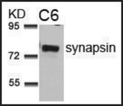
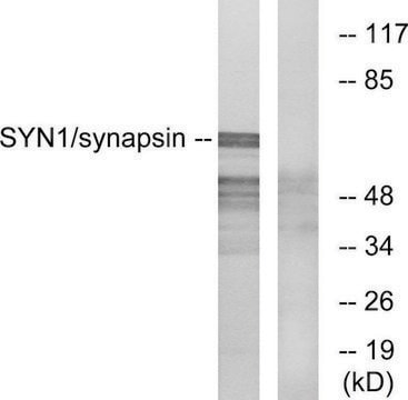
antigen ~80 kDa (Synapsin Ia)

species reactivity

rat, mouse, human, bovine

technique(s)

dot blot: 1:1000
immunohistochemistry (frozen sections): 1:2000
immunoprecipitation (IP): 1 μg
indirect immunofluorescence: 1:2000
western blot: 1:1000

UniProt accession no.

storage temp.

−20°C

target post-translational modification

unmodified

Gene Information

human ... SYN1(6853)
mouse ... Syn1(20964)
rat ... Syn1(24949)

General description

Synapsin I (1a and 1b) refers to two nearly identical phosphoproteins found in the cytoplasmic surface of synaptic vesicles of the CNS (central nervous system) and PNS(peripheral nervous system). It may be used to localize and detect synapsin I (synapsins Ia and Ib are collectively referred to as synapsin I) in nerve terminals.The SYN1 gene is mapped to human chromosome Xp11.3-p11.23.

Immunogen

synapsin I from bovine brain.

Application

Applications in which this antibody has been used successfully, and the associated peer-reviewed papers, are given below.


Rabbit polyclonal anti-Synapsin I antibody can be used for the localization and detection of synapsin I (synapsins Ia and Ib, approximately 80 kDa and 77 kDa, are collectively referred to as synapsin I) in nerve terminals.

Biochem/physiol Actions

Synapsin I is involved in the regulation of axonogenesis and synaptogenesis wherein it serves as a substrate of various protein kinases including those activated by cAMP, calciuim/calmodulin, mitogens, and cyclins. The antibody strongly reacts with the synapsin I doublet. Synapsin I is associated with vesicle trafficking. Mutation in this gene is one of the known cause for familial epilepsy and autism spectrum disorders.

Physical form

Lyophilized from ammonium bicarbonate (5 mM)

Disclaimer

Unless otherwise stated in our catalog or other company documentation accompanying the product(s), our products are intended for research use only and are not to be used for any other purpose, which includes but is not limited to, unauthorized commercial uses, in vitro diagnostic uses, ex vivo or in vivo therapeutic uses or any type of consumption or application to humans or animals.

Ještě jste nenalezli správný produkt?  

Vyzkoušejte náš produkt Nástroj pro výběr produktů.

Storage Class

13 - Non Combustible Solids

wgk_germany

WGK 2

flash_point_f

Not applicable

flash_point_c

Not applicable

ppe

Eyeshields, Gloves, type N95 (US)


Osvědčení o analýze (COA)

Vyhledejte osvědčení Osvědčení o analýze (COA) zadáním čísla šarže/dávky těchto produktů. Čísla šarže a dávky lze nalézt na štítku produktu za slovy „Lot“ nebo „Batch“.

Již tento produkt vlastníte?

Dokumenty související s produkty, které jste v minulosti zakoupili, byly za účelem usnadnění shromážděny ve vaší Knihovně dokumentů.

Navštívit knihovnu dokumentů

DNA hypomethylation of Synapsin II CpG islands associates with increased gene expression in bipolar disorder and major depression.
Cruceanu C, et al.
BMC Psychiatry, 16(1), 286-286 (2016)
Synapsin I (protein I), a nerve terminal-specific phosphoprotein. III. Its association with synaptic vesicles studied in a highly purified synaptic vesicle preparation.
Huttner W B, et al.
The Journal of Cell Biology, 96(5), 1374-1388 (1983)
PRICKLE1 interaction with SYNAPSIN I reveals a role in autism spectrum disorders.
Paemka L, et al.
PLoS ONE, 8(12), e80737-e80737 (2013)
Andrew San Antonio et al.
The Journal of comparative neurology, 522(6), 1333-1354 (2013-10-30)
The hippocampal CA2 subfield was initially identified by Lorente de Nó as an anatomically distinct region based on its cytoarchitectural features. Although there is an enormous body of literature on other hippocampal subfields (CA1 and CA3), relatively little is known
Nonsense-mediated mRNA decay and loss-of-function of the protein underlie the X-linked epilepsy associated with the W356? mutation in synapsin I.
Giannandrea M, et al.
PLoS ONE, 8(6), e67724-e67724 (2013)

Náš tým vědeckých pracovníků má zkušenosti ve všech oblastech výzkumu, včetně přírodních věd, materiálových věd, chemické syntézy, chromatografie, analytiky a mnoha dalších..

Obraťte se na technický servis.