Přejít k obsahu
Merck
Všechny fotografie(2)

Key Documents

E4156

Sigma-Aldrich

Anti-Early Endosomal Antigen 1 (N-terminal) antibody produced in rabbit

~1 mg/mL, affinity isolated antibody, buffered aqueous solution

Synonyma:

Anti-EEA1, Anti-Endosome-associated Protein p162, Anti-Zinc Finger FYVE Domain-containing Protein 2

Přihlásitk zobrazení cen stanovených pro organizaci a smluvních cen


About This Item

MDL number:
UNSPSC Code:
12352203
NACRES:
NA.41

biological source

rabbit

conjugate

unconjugated

antibody form

affinity isolated antibody

antibody product type

primary antibodies

clone

polyclonal

form

buffered aqueous solution

mol wt

antigen ~160 kDa

species reactivity

mouse, human, rat

concentration

~1 mg/mL

technique(s)

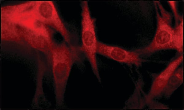
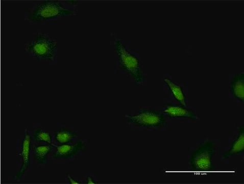
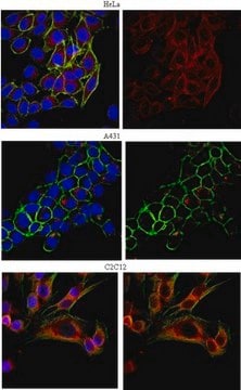
indirect immunofluorescence: 5-10 μg/mL using human HeLa and rat NRK cells
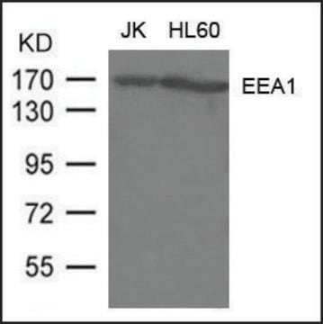
western blot (chemiluminescent): 0.4-0.8 μg/mL using whole extract of mouse NIH-3T3 cells

UniProt accession no.

shipped in

dry ice

storage temp.

−20°C

target post-translational modification

unmodified

Gene Information

human ... EEA1(8411)
mouse ... Eea1(216238)
rat ... Eea1(314764)

Související kategorie

General description

The gene Early Endosome Antigen 1 (EEA1) encodes for around 1400 amino acid proteins. It is a peripheral membrane protein associated with the cytoplasmic face of early endosomes. It is a 162 kDa autoantigen protein. EEA1 is a dimer, which comprises extensive coiled-coil regions. At its C-terminus, it contains a cysteine-rich zinc-finger-like domain named FYVE domain. This FYVE domain is conserved from yeast to man. FYVE domain is implicated in the specific localization of EEA1 to endosomes.

Immunogen

synthetic peptide corresponding amino acid residues 24-40 of human EEA1 with C-terminal added cysteine, conjugated to KLH. The corresponding sequence is identical in mouse.

Application

Anti-Early Endosomal Antigen 1 (N-terminal) antibody produced in rabbit has been used in immunoblotting and immunofluorescence.

Biochem/physiol Actions

Early Endosomal Antigen 1 (EEA1) localization in endosomes is implicated in subacute systemic lupus erythematosus. Endosomal targeting of EEA1 also requires its binding to the active form of the small GTPase Rab5. The binding of EEA1 to phosphatidylinositol 3 phosphate (PtdInsP) and rabaptin-5 (Rab5)-GTP is essential for the localization and function of EEA1 in endocytic membrane fusion. Anti-EEA1 may be used as an early endosome marker.

Physical form

Solution in 0.01 M phosphate buffered saline, pH 7.4, containing 15 mM sodium azide.

Disclaimer

Unless otherwise stated in our catalog or other company documentation accompanying the product(s), our products are intended for research use only and are not to be used for any other purpose, which includes but is not limited to, unauthorized commercial uses, in vitro diagnostic uses, ex vivo or in vivo therapeutic uses or any type of consumption or application to humans or animals.

Ještě jste nenalezli správný produkt?  

Vyzkoušejte náš produkt Nástroj pro výběr produktů.

Storage Class

10 - Combustible liquids

wgk_germany

WGK 3

flash_point_f

Not applicable

flash_point_c

Not applicable

ppe

Eyeshields, Gloves, multi-purpose combination respirator cartridge (US)


Osvědčení o analýze (COA)

Vyhledejte osvědčení Osvědčení o analýze (COA) zadáním čísla šarže/dávky těchto produktů. Čísla šarže a dávky lze nalézt na štítku produktu za slovy „Lot“ nebo „Batch“.

Již tento produkt vlastníte?

Dokumenty související s produkty, které jste v minulosti zakoupili, byly za účelem usnadnění shromážděny ve vaší Knihovně dokumentů.

Navštívit knihovnu dokumentů

FYVE and coiled-coil domains determine the specific localisation of Hrs to early endosomes
Raiborg C, et al.
Journal of Cell Science, 114(12), 2255-2263 (2001)
Stefanie Hetzenecker et al.
Traffic (Copenhagen, Denmark), 17(4), 351-368 (2015-12-10)
Human cytomegalovirus (HCMV) is an important and widespread pathogen in the human population. While infection by this β-herpesvirus in endothelial, epithelial and dendritic cells depends on endocytosis, its entry into fibroblasts is thought to occur by direct fusion of the
Distinct lysosomal network protein profiles in Parkinsonian syndrome cerebrospinal fluid
Boman A, et al.
Journal of Parkinson's Disease, 6(2), 307-315 (2016)
Andrea Boman et al.
Journal of Parkinson's disease, 6(2), 307-315 (2016-04-12)
Clinical diagnosis of parkinsonian syndromes like Parkinson's disease (PD), corticobasal degeneration (CBD) and progressive supranuclear palsy (PSP) is hampered by overlapping symptomatology and lack of diagnostic biomarkers, and definitive diagnosis is only possible post-mortem. Since impaired protein degradation plays an
Lipid interaction networks of peripheral membrane proteins revealed by data-driven micelle docking
Dancea F, et al.
Biophysical Journal, 94(2), 515-524 (2008)

Náš tým vědeckých pracovníků má zkušenosti ve všech oblastech výzkumu, včetně přírodních věd, materiálových věd, chemické syntézy, chromatografie, analytiky a mnoha dalších..

Obraťte se na technický servis.