
ABN1698
Anti-Aspa/Nur7 Antibody
from rabbit, purified by affinity chromatography
Synonyma:
Aspartoacylase, Aminoacylase-2, ACY-2, Aspa/Nur7, Anti-Aspartoacylase
Vybrat velikost
| Velikost balení | Skladová položka | Dostupnost | Cena |
|---|---|---|---|
| 100 μg | Očekávané datum odeslání10. června 2026odAreál Kühne+Nagel spol. s r.o. | 10 500,00 Kč |
O této položce
10 500,00 Kč
Očekávané datum odeslání10. června 2026podrobné informace
biological source
rabbit
Quality Segment
conjugate
unconjugated
antibody form
purified antibody
antibody product type
primary antibodies
clone
polyclonal
purified by
affinity chromatography
species reactivity
human, mouse, rat
technique(s)
immunohistochemistry: suitable, western blot: suitable
NCBI accession no.
UniProt accession no.
shipped in
wet ice
target post-translational modification
unmodified
Gene Information
human ... ASPA(443)
General description
Immunogen
Application
Western Blotting Analysis: A representative lot detected Aspa/Nur7 in the cytosol, but not myelin, fraction from rat brain tissue homogenate (Madhavarao, C.N., et al. (2004). J Comp Neurol. 472(3):318-329).
Immunohistochemistry Analysis: A representative lot detected Aspa/Nur7 expression pattern in various regions of rat forebrain, including corpus callosum, cerebral cortex, hippocampal commissure (hc), fimbria, and anterior commissure (Madhavarao, C.N., et al. (2004). J Comp Neurol. 472(3):318-329).
Immunohistochemistry Analysis: A representative lot detected similar Aspa/Nur7 expression pattern as CC1 in various regions of rat forebrain, including the Purkinjie & axonal fiber layers of cerebellum, corpus callosum, as well as layer 2 of primary somatosensory cortex (Madhavarao, C.N., et al. (2004). J Comp Neurol. 472(3):318-329).
Neuroscience
Developmental Signaling
Physical form
Preparation Note
Analysis Note
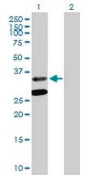
Western Blotting Analysis: 1.0 µg/mL of this antibody detected Aspa/Nur7 in 10 µg of rat brain tissue lysate.
Other Notes
Disclaimer
1 of 1
Tato položka | |||
|---|---|---|---|
| Quality Level 100 | Quality Level 100 | Quality Level 100 | Quality Level - |
| antibody form purified antibody | antibody form purified immunoglobulin | antibody form purified immunoglobulin | antibody form purified immunoglobulin |
| conjugate unconjugated | conjugate unconjugated | conjugate unconjugated | conjugate unconjugated |
| biological source rabbit | biological source mouse | biological source mouse | biological source rabbit |
| clone polyclonal | clone 3C11, monoclonal | clone polyclonal | clone polyclonal |
| UniProt accession no. | UniProt accession no. | UniProt accession no. | UniProt accession no. |


Still not finding the right product?
Vyzkoušejte náš nástroj Nástroj pro výběr produktů a zúžte své možnosti.
Skladovací třída
12 - Non Combustible Liquids
wgk
WGK 1
flash_point_f
Not applicable
flash_point_c
Not applicable
Osvědčení o analýze (COA)
Vyhledejte osvědčení Osvědčení o analýze (COA) zadáním čísla šarže/dávky těchto produktů. Čísla šarže a dávky lze nalézt na štítku produktu za slovy „Lot“ nebo „Batch“.
Již tento produkt vlastníte?
Dokumenty související s produkty, které jste v minulosti zakoupili, byly za účelem usnadnění shromážděny ve vaší Knihovně dokumentů.
Související obsah
A major focus of breast cancer research is to understand the mechanisms responsible for disease progression and drug resistance. Toward that end, it has been found that approximately two thirds of all human breast carcinomas overexpress the Estrogen Receptor α (ERα) protein and it remains the primary pharmacological target for endocrine therapy1,2. The normal cellular function of ERα is as a transcription factor that mediates a wide variety of physiological processes, many of which are dependent upon phosphorylation of the receptor at specific amino acid residues3,4. Indeed, ERα is known to be phosphorylated at a multitude of different sites, yet how these all correlate to disease remains unclear5. Here, we interrogated multiple sites of ERα for phosphorylation status by screening an extensive panel of different breast cancer patient samples and other non-breast cancer tissue microarray (TMA) slide samples to determine their relevance to disease.
Globální číslo obchodní položky
| Skladová položka | GTIN |
|---|---|
| ABN1698 | 04055977182569 |