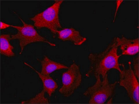
MAB3430
Anti-Desmin Antibody, clone DE-B-5
clone DE-B-5, Chemicon®, from mouse
About This Item
Productos recomendados
biological source
mouse
Quality Level
antibody form
purified antibody
clone
DE-B-5, monoclonal
species reactivity
mouse, frog, pig, rat, human
manufacturer/tradename
Chemicon®
technique(s)
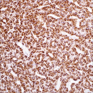
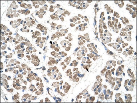
immunohistochemistry (formalin-fixed, paraffin-embedded sections): suitable
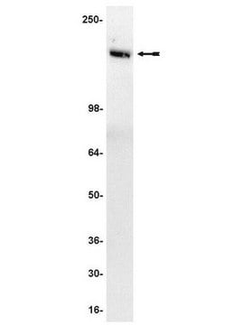
western blot: suitable
isotype
IgG1
NCBI accession no.
UniProt accession no.
shipped in
wet ice
target post-translational modification
unmodified
Gene Information
human ... DES(1674)
Specificity
Immunogen
Application
Optimal working dilutions must be determined by end user.
Immunohysto/cyto chemistry Protocols
Ideal specimens are obtained from frozen sections from shock-frozen tissue samples. The frozen sections are dried in the air and then fixed with acetone at -20°C for 10 min. Excess acetone is allowed to evaporate at 15-25°C. Material fixed in alcohol and embedded in paraffin can also be used (2). Formaldehyde fixation will reduce or eliminate the intensity of staining depending on the conditions under which it is performed. Other fixation conditions must be first tested by the investigator.
It is advantageous to block unspecific binding sites by overlaying the sections with fetal calf serum for 20-30 min at 15-25°C. Excess of fetal calf serum is removed by decanting before application of the antibody solution.
Cytocentrifuge preparations of single cells or cell smears are also fixed in acetone. These preparations should, however not be dried in the air. Instead, the excess acetone is removed by briefly washing in phosphate-buffered saline (PBS).
Further treatment is then as follows:
• Overlay the preparation with 10-20 μl antibody solution and incubate in a humid chamber at 37°C for 1 h.
• Dip the slide briefly in PBS and then wash 3 x in PBS for 3 min (using a fresh PBS bath in each case).
• Wipe the margins of the preparation dry and overlay the preparation with 10-20 μl of a solution of anti-mouse Ig-FITC or anti-mouse IgG-peroxidase solution and allow to incubate for 1 h at 37°C in a humid chamber.
• Wash the slide as described above.
The preparation must not be allowed to dry out during any of the steps.
If using an indirect immunofluorescence technique, the preparation should be overlaid with a suitable embedding medium (e.g. Moviol, Hoechst) and examined under the fluorescence microscope. If a POD-conjugate has been used as the secondary antibody, the preparation should be overlaid with a substrate solution (see below) and incubated at 15-25°C until a clearly visible redbrown color develops. A negative control (e.g. only the secondary antibody) should remain unchanged in color during this incubation period. Subsequently, the substrate is washed off with PBS and the preparation is stained, if desired, with hemalum stain for about 1 min. The hemalum solution is washed off with PBS; the preparation is embedded and examined.
Substrate solutions:
Aminoethyl-carbazole: Dissolve 2 mg 3-amino-9-ethylcarbazole with 1.2 ml dimethylsulfoxide and add 28.8 ml 50 mM Tris-HCI, pH 7.3, and 20 μl 3% H 2 O 2 (w/v). Prepare solution freshly each day. Diaminobenzidine: Dissolve 25 mg 3,3′-diaminobenzidine with 50 ml 50 mM Tris-HCI, pH 7.3, and add 40 μl 3% H 2 O 2 (w/v). Prepare solution freshly each day.
Cell Structure
Cytoskeleton
Quality
Linkage
Physical form
Storage and Stability
Other Notes
Legal Information
Disclaimer
Optional
Storage Class
12 - Non Combustible Liquids
wgk_germany
WGK 2
flash_point_f
Not applicable
flash_point_c
Not applicable
Certificados de análisis (COA)
Busque Certificados de análisis (COA) introduciendo el número de lote del producto. Los números de lote se encuentran en la etiqueta del producto después de las palabras «Lot» o «Batch»
¿Ya tiene este producto?
Encuentre la documentación para los productos que ha comprado recientemente en la Biblioteca de documentos.
Nuestro equipo de científicos tiene experiencia en todas las áreas de investigación: Ciencias de la vida, Ciencia de los materiales, Síntesis química, Cromatografía, Analítica y muchas otras.
Póngase en contacto con el Servicio técnico