
11296736001
Roche
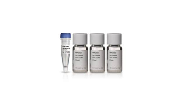
5-Bromo-2′-deoxy-uridine Labeling and Detection Kit I
sufficient for ≤100 tests, kit of 1 (5 components), suitable for immunofluorescence
Synonyme(s) :
5-BrdU, 5-Bromo-2-deoxyuridine
About This Item
Produits recommandés


Utilisation
sufficient for ≤100 tests
Niveau de qualité
Conditionnement
kit of 1 (5 components)
Fabricant/nom de marque
Roche
Technique(s)
immunofluorescence: suitable
Température de stockage
−20°C
Description générale
Normally, binding of the antibody is only achieved by denaturation of the DNA. This is usually obtained by exposing the cells to acid, base, or heat. These procedures result in destruction of cell integrity, including cell morphology and surface and cytoplasmatic markers.
The BrdU Labeling and Detection Kit I avoids these problems. The antibody preparation contains specific nucleases which allows access to BrdU after fixation in acidic ethanol. Therefore also simultaneous detection of other markers (double staining) is possible.
Spécificité
Application
- Safe: No radioisotopes are used
- Easy to perform: Follows a standard immunofluorescence protocol
- Sensitive: Denaturation of DNA with nucleases allows for highly sensitive detection of BrdU
- Flexible: Allows double-labeling protocols
BrdU Labeling and Detection Kit has been used for the detection of 5-bromo-2′-deoxy-uridine (BrdU) incorporated into cellular DNA.
Conditionnement
Notes préparatoires
Working solution: BrdU labeling medium
Dilute BrdU labeling reagent 1:1000 with sterile cell culture medium (final concentration 10μM).
Note: For in vivo labeling undiluted BrdU labeling reagent (1 to 2ml/100 g body weight) is needed.
Prepare shortly before use.
Anti-BrdU working solution
Dilute anti-BrdU solution 1:10 with Incubation buffer.
Prepare shortly before use.
Anti-mouse-Ig-fluorescein stock solution
Dissolve anti-mouse-Ig-fluorescein solution in 1ml double-dist. water.
Anti-mouse-Ig-fluorescein working solution
Dilute anti-mouse Ig-fluorescein stock solution 1:10 with PBS. If an extended storage is desired, add BSA (bovine serum albumin), 10 mg/ml.
Prepare shortly before use.
Washing buffer
Dilute Washing buffer concentrate (10x) (bottle 2) 1:10 with double-dist. water.
Storage conditions (working solution): BrdU labeling medium
Store undiluted (1000x) medium in aliquots at -15 to -25°C.
Anti-BrdU working solution
Store undiluted antibody at -15 to -25°C.
Anti-mouse-Ig-fluorescein stock solution
Stable at 2 to 8°C
Washing buffer
Stable at 2 to 8°C
Sample material: Cell culture: adherent cells, suspension cells, organ, or explant cultures. Tissue sections (after in vivo labeling with BrdU).
Autres remarques
Composants de kit seuls
- BrdU Labeling Reagent, sterile 1,000x concentrated
- Washing Buffer concentrate 10x concentrated
- Incubation Buffer
- Anti-BrdU antibody, contains nucleases for DNA denaturation
- Anti-mouse-Ig-fluorescein antibody


Mention d'avertissement
Danger
Mentions de danger
Conseils de prudence
Classification des risques
Aquatic Chronic 3 - Eye Irrit. 2 - Muta. 1B - Skin Irrit. 2 - Skin Sens. 1
Code de la classe de stockage
12 - Non Combustible Liquids
Classe de danger pour l'eau (WGK)
WGK 2
Point d'éclair (°F)
does not flash
Point d'éclair (°C)
does not flash
Faites votre choix parmi les versions les plus récentes :
Déjà en possession de ce produit ?
Retrouvez la documentation relative aux produits que vous avez récemment achetés dans la Bibliothèque de documents.
Les clients ont également consulté


Articles
Tests sur cellules pour l'étude de la prolifération (BrdU, MTT, WST1), de la viabilité et de la toxicité cellulaires pour la recherche sur le cancer, la recherche en neurosciences et la recherche sur cellules souches.
Cell based assays for cell proliferation (BrdU, MTT, WST1), cell viability and cytotoxicity experiments for applications in cancer, neuroscience and stem cell research.
Cell based assays for cell proliferation (BrdU, MTT, WST1), cell viability and cytotoxicity experiments for applications in cancer, neuroscience and stem cell research.
Cell based assays for cell proliferation (BrdU, MTT, WST1), cell viability and cytotoxicity experiments for applications in cancer, neuroscience and stem cell research.
Notre équipe de scientifiques dispose d'une expérience dans tous les secteurs de la recherche, notamment en sciences de la vie, science des matériaux, synthèse chimique, chromatographie, analyse et dans de nombreux autres domaines..
Contacter notre Service technique