
AB2047
Anti-Fibronectin Antibody
Chemicon®, from rabbit
About This Item
Produits recommandés


Source biologique
rabbit
Forme d'anticorps
affinity purified immunoglobulin
Type de produit anticorps
primary antibodies
Clone
polyclonal
Espèces réactives
bovine
Fabricant/nom de marque
Chemicon®
Technique(s)
ELISA: suitable
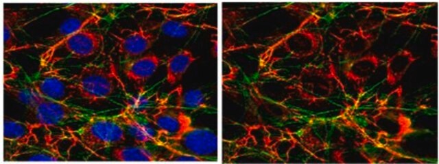
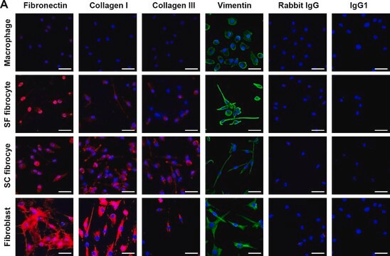
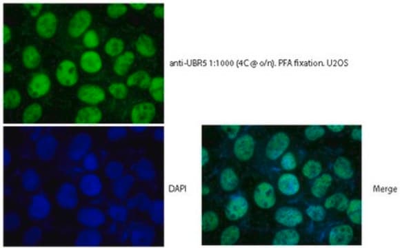
immunofluorescence: suitable
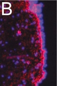
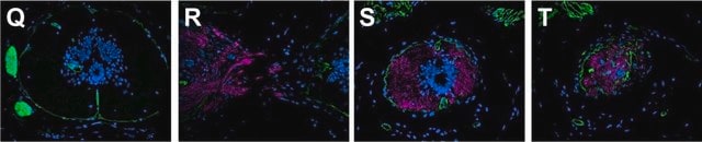
immunohistochemistry: suitable (paraffin)
radioimmunoassay: suitable
western blot: suitable
Numéro d'accès UniProt
Conditions d'expédition
dry ice
Modification post-traductionnelle de la cible
unmodified
Description générale
Fibronectin exists in two main forms: 1) as an insoluble glycoprotein dimer that serves as a linker in the ECM (extracellular matrix), and; 2) as a soluble disulphide linked dimer found in the plasma (plasma FN). The plasma form is synthesized by hepatocytes, and the ECM form is made by fibroblasts, chondrocytes, endothelial cells, macrophages, as well as certain epithelial cells.
Fibronectin sometimes serves as a general cell adhesion molecule by anchoring cells to collagen or proteoglycan substrates. FN also can serve to organize cellular interaction with the ECM by binding to different components of the extracellular matrix and to membrane-bound FN receptors on cell surfaces. The importance of fibronectin in cell migration events during embryogenesis has been documented in several contexts, e.g.: 1) mesodermal cell migration during gastrulation can be blocked by injection of Arg-Gly-Asp (RGD) tripeptides that block cellular FN receptors (integrins); 2) injection of anti-FN antibodies into chick embryos blocks migration of precardiac cells to the embryonic midline, and; 3) the patterns of FN deposition in developing vertebrate limbs determines the patterns of precartilage cell adhesion to the ECM, thereby specifying limb-specific patterns of chondrogenesis. {D. Marcey, http://www.clunet.edu/BioDev/omm/fibro/frames/fibrotxt.htm}.
Spécificité
Immunogène
Application
Immunohistochemistry on paraffin embedded tissues requires light fixation in 2% PFA, 4% formalin (less than 90 minutes), acetone or methyl-carnoy fixation; traditional formalin fixation is not recommended. Antigen retrieval is HIER citrate buffer; detection is via enhanced enzymatic methods only.
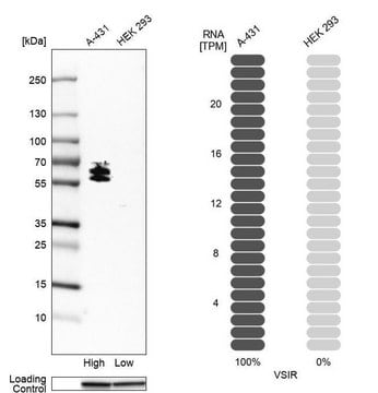
Immunoblotting: 1:1000 of 2% deoxycholate + 10% SDS, 6M urea extracted bovine cell cultures (Kinsella, 2000). Antibody demonstates the appropriate twin bands at ~220kDa.
Radioimmunoassay
ELISA
Optimal working dilutions must be determined by end user.
Cell Structure
ECM Proteins
Forme physique
Stockage et stabilité
Autres remarques
Informations légales
Clause de non-responsabilité
Vous ne trouvez pas le bon produit ?
Essayez notre Outil de sélection de produits.
Code de la classe de stockage
12 - Non Combustible Liquids
Classe de danger pour l'eau (WGK)
WGK 2
Point d'éclair (°F)
Not applicable
Point d'éclair (°C)
Not applicable
Certificats d'analyse (COA)
Recherchez un Certificats d'analyse (COA) en saisissant le numéro de lot du produit. Les numéros de lot figurent sur l'étiquette du produit après les mots "Lot" ou "Batch".
Déjà en possession de ce produit ?
Retrouvez la documentation relative aux produits que vous avez récemment achetés dans la Bibliothèque de documents.
Active Filters
Notre équipe de scientifiques dispose d'une expérience dans tous les secteurs de la recherche, notamment en sciences de la vie, science des matériaux, synthèse chimique, chromatographie, analyse et dans de nombreux autres domaines..
Contacter notre Service technique