Pular para o conteúdo
Merck
Todas as fotos(1)

Documentos Principais

MABT855M

Sigma-Aldrich

Anti-Podoplanin Antibody, clone LpMab-13

clone LpMab-13, from mouse

Sinônimo(s):

Aggrus, Glycoprotein 36, Gp36, PA2.26 antigen, T1-alpha, T1A

Faça loginpara ver os preços organizacionais e de contrato


About This Item

Código UNSPSC:
12352203
eCl@ss:
32160702
NACRES:
NA.41
Preço e disponibilidade não estão disponíveis no momento.

fonte biológica

mouse

Nível de qualidade

forma do anticorpo

purified immunoglobulin

tipo de produto de anticorpo

primary antibodies

clone

LpMab-13, monoclonal

reatividade de espécies

human

embalagem

antibody small pack of 25 μg

técnica(s)

ELISA: suitable
flow cytometry: suitable
immunohistochemistry: suitable (paraffin)
western blot: suitable

Isotipo

IgG1κ

nº de adesão NCBI

nº de adesão UniProt

modificação pós-traducional do alvo

unmodified

Informações sobre genes

human ... PDPN(10630)

Descrição geral

Podoplanin (UniProt: Q86YL7; also known as Aggrus, Glycoprotein 36, Gp36, PA2.26 antigen, T1-alpha, T1A) is encoded by the PDPN (also known as GP36) gene (Gene ID: 10630) in human. Podoplanin serves as the endogenous ligand of C-type lectin-like receptor-2 (CLEC-2) and is highly expressed in various tumors and in some normal cells, such as lymphatic endothelial cells and podocytes. It may be involved in cell migration and/or actin cytoskeleton organization. Podoplanin is localized to actin-rich microvilli and plasma membrane projections, such as filopodia, lamellipodia, and ruffles. Six isoforms of podoplanin have been described that are produced by alternative splicing. It is synthesized with a signal peptide of 22 amino acids, which is cleaved to produce the mature form. Podoplanin contains an extracellular domain (aa 23-131), a helical domain (aa 132-152) and a cytoplasmic region (aa 153-162). Podoplanin possesses three tandem repeat of platelet aggregation-stimulating (PLAG) domains in its N-terminus. Among the PLAG domains, sialylated O-glycan on Thr52 of PLAG3 is essential for the binding to C-type lectin-like receptor-2 (CLEC-2). Interaction of human podoplanin with CLEC-2 mainly involves Glu47 and Asp48 in the platelet PLAG3. Clone LpMab-13 binds to PLAG2/3 domains and recognizes endogenous hPDPN of cancer cells, including glioblastoma, oral cancer, lung cancer, and malignant mesothelioma, and normal cells such as lymphatic endothelial cells and podocytes of kidney. The interaction between LpMab-13 and hPDPN is independent of its glycosylation. The minimum epitope of LpMab-13 is identified to be Ala42-Asp49 of hPDPN. ((Ref.: Ogasawara, S., et al. (2016). Monoclon. Antib. immunodiag. Immunother. 35(3); 155-163).

Especificidade

Clone LpMab-13 detects Podoplanin in human cells. The minimal epitope is shown to be Ala42-Asp49.

Imunogênio

LN229 glioma cells expressing human Podoplanin.

Aplicação

Anti-Podoplanin, clone LpMab-13, Cat. No. MABT855, is a mouse monoclonal antibody that detects human podoplanin and has been tested for use in ELISA, Flow Cytometry, Immunohistochemistry (Paraffin), and Western Blotting.
ELISA Analysis: A representative lot detected Podoplanin in ELISA application (Ogasawara, S., et. al. (2016). Monoclon Antib Immunodiagn Immunother. 35(3):155-62).

Flow Cytometry Analysis: A representative lot detected Podoplanin in Flow Cytometry application (Ogasawara, S., et. al. (2016). Monoclon Antib Immunodiagn Immunother. 35(3):155-62).

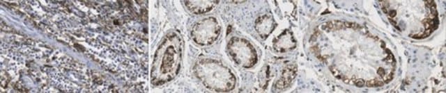
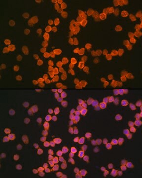
Immunohistochemistry Analysis: A representative lot detected Podoplanin in Immunohistochemistry application (Ogasawara, S., et. al. (2016). Monoclon Antib Immunodiagn Immunother. 35(3):155-62).

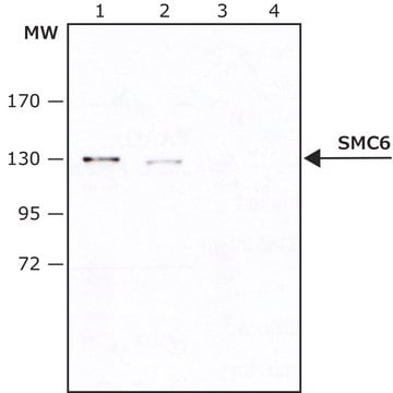
Western Blotting Analysis: A representative lot detected Podoplanin in Western Blotting application (Ogasawara, S., et. al. (2016). Monoclon Antib Immunodiagn Immunother. 35(3):155-62).
Research Category
Cell Structure

Qualidade

Evaluated by Immunohistochemistry in human testis cancer tissue.

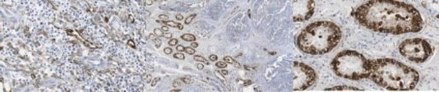
Immunohistochemistry Analysis: A 1:250 dilution of this antibody detected Podoplanin in human testis cancer tissue.

Descrição-alvo

16.70 kda calculated.

forma física

Format: Purified
Protein G purified
Purified mouse monoclonal antibody IgG1 in buffer containing 0.1 M Tris-Glycine (pH 7.4), 150 mM NaCl with 0.05% sodium azide.

Armazenamento e estabilidade

Stable for 1 year at 2-8°C from date of receipt.

Outras notas

Concentration: Please refer to lot specific datasheet.

Exoneração de responsabilidade

Unless otherwise stated in our catalog or other company documentation accompanying the product(s), our products are intended for research use only and are not to be used for any other purpose, which includes but is not limited to, unauthorized commercial uses, in vitro diagnostic uses, ex vivo or in vivo therapeutic uses or any type of consumption or application to humans or animals.

Não está encontrando o produto certo?  

Experimente o nosso Ferramenta de seleção de produtos.

Código de classe de armazenamento

12 - Non Combustible Liquids

Classe de risco de água (WGK)

WGK 1

Ponto de fulgor (°F)

Not applicable

Ponto de fulgor (°C)

Not applicable


Certificados de análise (COA)

Busque Certificados de análise (COA) digitando o Número do Lote do produto. Os números de lote e remessa podem ser encontrados no rótulo de um produto após a palavra “Lot” ou “Batch”.

Já possui este produto?

Encontre a documentação dos produtos que você adquiriu recentemente na biblioteca de documentos.

Visite a Biblioteca de Documentos

Questions

Reviews

No rating value

Active Filters

Nossa equipe de cientistas tem experiência em todas as áreas de pesquisa, incluindo Life Sciences, ciência de materiais, síntese química, cromatografia, química analítica e muitas outras.

Entre em contato com a assistência técnica