MABS1249
Anti-SCAP Antibody, clone 9D5
clone 9D5, from mouse
Sinônimo(s):
Sterol regulatory element-binding protein cleavage-activating protein, SCAP, SREBP cleavage-activating protein
About This Item
Produtos recomendados
fonte biológica
mouse
Nível de qualidade
forma do anticorpo
purified immunoglobulin
tipo de produto de anticorpo
primary antibodies
clone
9D5, monoclonal
reatividade de espécies
hamster
técnica(s)
immunocytochemistry: suitable
western blot: suitable
Isotipo
IgG2bκ
nº de adesão NCBI
nº de adesão UniProt
modificação pós-traducional do alvo
unmodified
Informações sobre genes
hamster ... Scap(100689048)
Descrição geral
4 lumenal and 5 cytoplasmic regions, having both its N- and C-terminal ends at the cytoplasmic side (a.a. 1-18 & 730-1276). The C-terminal domain of SCAP mediates association with SREBPs, while transmembrane helices 2–6 comprise the sterol-sensing domain that mediates sterol-induced binding of SCAP to Insigs. Mutations within the sterol-sensing region disrupt Insig binding and prevent sterol-mediated ER retention of SCAP-SREBP.
Especificidade
Imunogênio
Aplicação
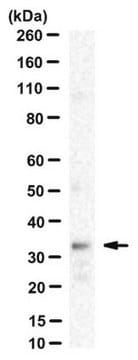
Western Blotting Analysis: A representative lot detected exogenously expressed SCAP co-immunoprecipitated with the exogenously expressed Insig-1 and Insig-2 upon 25-hydroxycholesterol (25-HC) treatment of a SCAP-deficient CHO cell line SRD-13A that had been transfected to co-express SCAP with either myc-tagged Insig-1 or Insig-2 (Lee, P.C., and DeBose-Boyd, R.A. (2010). J. Lipid Res. 51(1):192-201).
Western Blotting Analysis: Representative lots detected similar level of SCAP in the membrane extracts from CHO cells with or without SREBP processing inhibitor 25-hydroxycholesterol (25-HC) treatment regardless whether the cells′ 25-HC-resistant status (Lee, P.C., and DeBose-Boyd, R.A. (2010). J. Lipid Res. 51(1):192-201; Lee, P.C., et al. (2007). J. Lipid Res. 48(9):1944-1954).
Western Blotting Analysis: A representative lot detected the endogenous SCAP immunoprecipitated from CHO cell lysate by a polyclonal SCAP antibody (Sakai, J., et al. (1997). J. Biol. Chem. 272(32):20213-20221).
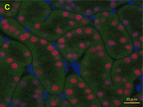
Immunocytochemistry Analysis: A representative lot localized the overexpressed hamster SCAP at ER and nuclear envelope by fluorescent immunocytochemistry staining of 3% paraformaldehyde-fixed, 0.01% saponin-permeabilized CHO stable transfectants (Sakai, J., et al. (1997). J. Biol. Chem. 272(32):20213-20221).
Qualidade
Isotyping Analysis: The identity of this monoclonal antibody is confirmed by isotyping test to be IgG2bκ.
Descrição-alvo
forma física
Outras notas
Not finding the right product?
Try our Ferramenta de seleção de produtos.
Código de classe de armazenamento
12 - Non Combustible Liquids
Classe de risco de água (WGK)
WGK 1
Ponto de fulgor (°F)
Not applicable
Ponto de fulgor (°C)
Not applicable
Certificados de análise (COA)
Busque Certificados de análise (COA) digitando o Número do Lote do produto. Os números de lote e remessa podem ser encontrados no rótulo de um produto após a palavra “Lot” ou “Batch”.
Já possui este produto?
Encontre a documentação dos produtos que você adquiriu recentemente na biblioteca de documentos.
Nossa equipe de cientistas tem experiência em todas as áreas de pesquisa, incluindo Life Sciences, ciência de materiais, síntese química, cromatografia, química analítica e muitas outras.
Entre em contato com a assistência técnica


