Pular para o conteúdo
Merck
Todas as fotos(1)

Documentos Principais

MABF939

Sigma-Aldrich

Anti-IFNAR1 Antibody, clone 4G8

clone 4G8, from mouse

Sinônimo(s):

Interferon alpha/beta receptor 1, CRF2-1, Cytokine receptor class-II member 1, Cytokine receptor family 2 member 1, IFN-alpha/beta receptor 1, IFN-R-1, Type I interferon receptor 1

Faça loginpara ver os preços organizacionais e de contrato


About This Item

Código UNSPSC:
12352203
eCl@ss:
32160702
Preço e disponibilidade não estão disponíveis no momento.

fonte biológica

mouse

Nível de qualidade

forma do anticorpo

purified immunoglobulin

tipo de produto de anticorpo

primary antibodies

clone

4G8, monoclonal

reatividade de espécies

human

técnica(s)

flow cytometry: suitable

Isotipo

IgG1κ

nº de adesão NCBI

nº de adesão UniProt

Condições de expedição

ambient

modificação pós-traducional do alvo

unmodified

Informações sobre genes

human ... IFNAR1(3454)

Categorias relacionadas

Descrição geral

Interferon alpha/beta receptor 1 (UniProt P17181; also known as CRF2-1, Cytokine receptor class-II member 1, Cytokine receptor family 2 member 1, IFN-alpha/beta receptor 1, IFN-R-1, Type I interferon receptor 1) is encoded by the IFNAR1 (also known as IFNAR) gene (Gene ID 3454) in human. Interferons (IFNs) are cytokines whose prouction and release by host cells are induced in the presence of pathogens (e.g. viruses, bacteria, parasites) and tumor cells. More than twenty distinct IFN genes and proteins have been identified and classified into three main types. Type I IFNs (IFN-α, IFN-β, IFN-ε, IFN-κ, and IFN-ω) exert their function via binding IFN-α/β receptor (encoded by the IFNAR1 and IFNAR2 gene, respectively). Ligation of IFNAR by IFN-I results in activation of the Janus kinases, Jak1 and Tyk2, which in turn phosphorylates STAT1 and STAT2, resulting in recruitment of IRF9 to form the IFN-stimulated gene factor 3 (ISGF3) transcription factor. ISGF3 then translocates to the nucleus, where it binds IFN-stimulated response elements (ISREs) and drives the transcription of IFN-stimulated genes (ISGs). IFNAR1 is phosphorylated by Tyk2 and PKD2 upon IFN-I stimulation, leading to IFNAR1 ubiquitination and lysosomal degradation. IFNAR1 is also
phosphorylated by p38 MAP kinase in response to non-IFN stimuli, including the PERK-dependent unfolded protein response (UPR), ligation of pattern recognition receptors (PRRs), or through signaling via other inflammatory cytokines or growth factors including VEGF, IL-1β and TNFα. Encephalitic flaviviruses antagonize IFN-I signaling by inhibiting IFNAR1 surface expression, where the viral nonstructural protein 5 (NS5) targets cellular prolidase (PEPD) that is required for IFNAR1 maturation and accumulation.

Especificidade

Clone 4G8 detected a loss of HEK293 cell surface IFNAR1 immunoreactivity following lentivirus-mediated cellular IFNAR1 shRNA delivery or West Nile virus (WNV) infection (Lubick, K.J., et al. (2015). Cell Host Microbe. 18(1):61-74).

Imunogênio

Epitope: extracellular domain
His-tagged recombinant human IFNAR1 extracellular domain.

Aplicação

Flow Cytometry Analysis: A representative lot detected a time-dependent loss of HEK293 cell surface IFNAR1 immunoreactivity following West Nile virus (WNV) infection. A significantly faster surface IFNAR1 loss was observed when cells were infected with WNV that produces IFN-I-antagonizing NS5 (nonstructural protein 5) with S653F mutation (Lubick, K.J., et al. (2015). Cell Host Microbe. 18(1):61-74).

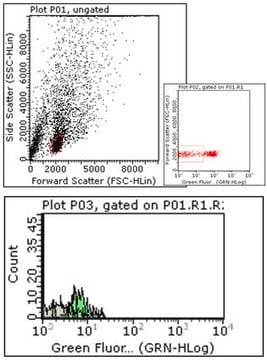
Flow Cytometry Analysis: A representative lot detected a loss of HEK293 cell surface IFNAR1 immunoreactivity following lentivirus-mediated cellular IFNAR1 shRNA delivery (Lubick, K.J., et al. (2015). Cell Host Microbe. 18(1):61-74).
Research Category
Inflammation & Immunology
This mouse monoclonal Anti-IFNAR1 Antibody, clone 4G8, Cat. No. MABF939 is validated for use in Flow Cytometry for the detection of IFNAR1.

Qualidade

Evaluated by Flow Cytometry in K562 cells.

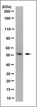
Flow Cytometry Analysis: 1 µg of this antibody detected IFNAR1 on the surface of K562 cells.

Descrição-alvo

63.53/60.93 kDa (pro-/mature isoform 1), 49.55/46.95 kDa (pro-/mature isoform 2), 56.72/54.12 kDa (pro-/mature isoform 3), 56.05 kDa (isoform 4) calculated. Apparent molecular weight appears greater than 100 kDa due to glycosylation.

forma física

Format: Purified
Protein G purified.
Purified mouse IgG1κ in buffer containing 0.1 M Tris-Glycine (pH 7.4), 150 mM NaCl with 0.05% sodium azide.

Armazenamento e estabilidade

Stable for 1 year at 2-8°C from date of receipt.

Outras notas

Concentration: Please refer to lot specific datasheet.

Exoneração de responsabilidade

Unless otherwise stated in our catalog or other company documentation accompanying the product(s), our products are intended for research use only and are not to be used for any other purpose, which includes but is not limited to, unauthorized commercial uses, in vitro diagnostic uses, ex vivo or in vivo therapeutic uses or any type of consumption or application to humans or animals.

Não está encontrando o produto certo?  

Experimente o nosso Ferramenta de seleção de produtos.

Código de classe de armazenamento

12 - Non Combustible Liquids

Classe de risco de água (WGK)

WGK 1

Ponto de fulgor (°F)

Not applicable

Ponto de fulgor (°C)

Not applicable


Certificados de análise (COA)

Busque Certificados de análise (COA) digitando o Número do Lote do produto. Os números de lote e remessa podem ser encontrados no rótulo de um produto após a palavra “Lot” ou “Batch”.

Já possui este produto?

Encontre a documentação dos produtos que você adquiriu recentemente na biblioteca de documentos.

Visite a Biblioteca de Documentos

Questions

Reviews

No rating value

Active Filters

Nossa equipe de cientistas tem experiência em todas as áreas de pesquisa, incluindo Life Sciences, ciência de materiais, síntese química, cromatografia, química analítica e muitas outras.

Entre em contato com a assistência técnica