MAB3839-I
Anti-Transglutaminase-2 Antibody, FN binding domain Antibody, clone 4G3
clone 4G3, from mouse
Synonyme(s) :
Protein-glutamine gamma-glutamyltransferase 2, EC: 2.3.2.13, Tissue transglutaminase, Transglutaminase C, TG(C), TGC, TGase C, Transglutaminase H, TGase H, TGase-2
About This Item
Produits recommandés
Source biologique
mouse
Forme d'anticorps
purified immunoglobulin
Type de produit anticorps
primary antibodies
Clone
4G3, monoclonal
Espèces réactives
human
Conditionnement
antibody small pack of 25 μg
Technique(s)
flow cytometry: suitable
immunofluorescence: suitable
immunoprecipitation (IP): suitable
western blot: suitable
Isotype
IgG1κ
Numéro d'accès NCBI
Numéro d'accès UniProt
Modification post-traductionnelle de la cible
unmodified
Informations sur le gène
human ... TGM2(7052)
Description générale
Spécificité
Immunogène
Application
Signaling
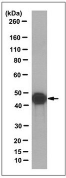
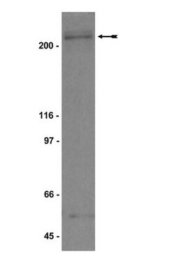
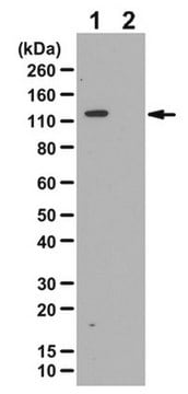
Western Blotting Analysis: A representative lot detected Transglutaminase-2, FN binding domain in Western Blotting applications (Janiak, A., et. al. (2006). Mol Biol Cell. 17(4):1606-19).
Radioimmunoassay Analysis: A representative lot detected Transglutaminase-2, FN binding domain in Radioimmunoassay applications (Akimov, S.S., et. al. (2001). Blood. 98(5):1567-76).
Flow Cytometry Analysis: A representative lot detected Transglutaminase-2, FN binding domain in Flow Cytometry applications (Zemskov, E.A., et. al. (2011). PLos One. 6(4):e19414).
Immunoprecipitation Analysis: A representative lot immunoprecipitated Transglutaminase-2, FN binding domain in Immunoprecipitation applications (Zemskov, E.A., et. al. (2011). PLos One. 6(4):e19414; Janiak, A., et. al. (2006). Mol Biol Cell. 17(4):1606-19).
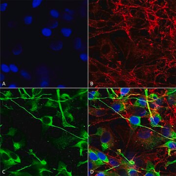
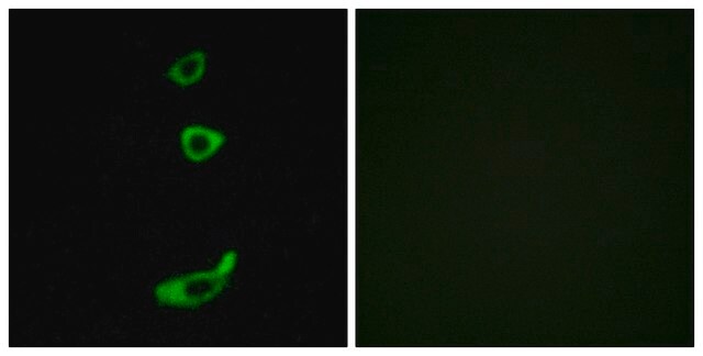
Immunofluorescence Analysis: A representative lot detected Transglutaminase-2, FN binding domain in Immunofluorescence applications (Janiak, A., et. al. (2006). Mol Biol Cell. 17(4):1606-19; Akimov, S.S., et. al. (2001). Blood. 98(5):1567-76; Zemskov, E.A., et. al. (2011). PLos One. 6(4):e19414).
Qualité
Western Blotting Analysis: 1 µg/mL of this antibody detected Transglutaminase-2, FN binding domain in A431 cell lysate.
Description de la cible
Liaison
Forme physique
Stockage et stabilité
Handling Recommendations: Upon receipt and prior to removing the cap, centrifuge the vial and gently mix the solution. Aliquot into microcentrifuge tubes and store at -20°C. Avoid repeated freeze/thaw cycles, which may damage IgG and affect product performance.
Autres remarques
Clause de non-responsabilité
Not finding the right product?
Try our Outil de sélection de produits.
Certificats d'analyse (COA)
Recherchez un Certificats d'analyse (COA) en saisissant le numéro de lot du produit. Les numéros de lot figurent sur l'étiquette du produit après les mots "Lot" ou "Batch".
Déjà en possession de ce produit ?
Retrouvez la documentation relative aux produits que vous avez récemment achetés dans la Bibliothèque de documents.
Notre équipe de scientifiques dispose d'une expérience dans tous les secteurs de la recherche, notamment en sciences de la vie, science des matériaux, synthèse chimique, chromatographie, analyse et dans de nombreux autres domaines..
Contacter notre Service technique