Saltar al contenido
Merck
Todas las fotos(1)

Documentos

MABF261

Sigma-Aldrich

Anti-PD-1 Antibody, clone 16A2.1

clone 16A2.1, from mouse, purified by affinity chromatography

Sinónimos:

PD-1, Programmed cell death protein 1, Protein PD-1, hPD-1, CD279

Iniciar sesiónpara Ver la Fijación de precios por contrato y de la organización


About This Item

UNSPSC Code:
12352203
eCl@ss:
32160702
NACRES:
NA.41

biological source

mouse

Quality Level

antibody form

purified immunoglobulin

antibody product type

primary antibodies

clone

16A2.1, monoclonal

purified by

affinity chromatography

species reactivity

human

technique(s)

flow cytometry: suitable
western blot: suitable

isotype

IgG2aκ

NCBI accession no.

UniProt accession no.

shipped in

wet ice

target post-translational modification

unmodified

Gene Information

human ... PDCD1(5133)

General description

Programmed cell death protein 1 (PD-1) (UniProt Q15116), also known as CD279, hPD-1, hPD-I, hSLE1, Protein PD-1, Systemic lupus erythematosus susceptibility 2) is a type I single transmembrane protein encoded by the PDCD1 gene in human. Maintenance of peripheral tolerance by preventing the development of autoimmunity due to T lymphocytes specific for self-antigens is essential for homeostasis of the immune system. PD-1 receptor CD279 and its ligands PD-L1 (B7-H1, CD274) and PD-L2 (B7-DC; CD273) play a vital role in the induction and maintenance of peripheral tolerance. Upregulated PD-1 expression is seen on conventional CD4+ CD25- T cells (Tconv) when exposed to mesenchymal stem cells, resulting in increased Tconv apoptosis due to PD-1 stimulation by the MSC PD-L2.

Immunogen

Epitope: Extracellular domain
KLH-conjugated linear peptide corresponding to the extracellular domain of human PD-1.

Application

Anti-PD-1 Antibody, clone 16A2.1 is an antibody against PD-1 for use in Western Blotting, Flow Cytometry.
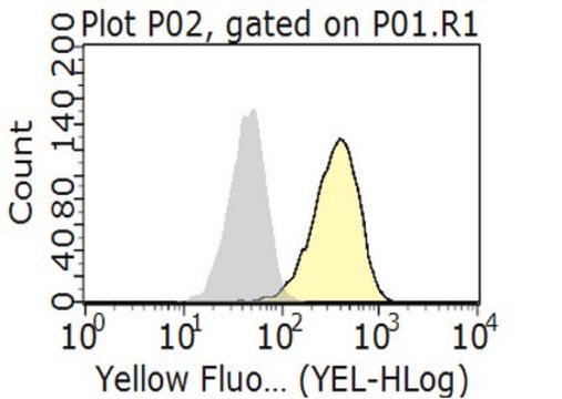
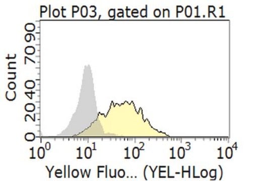
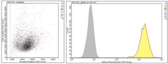
Flow Cytometry Analysis: 2.0 µg of this antibody from a representative lot detected PD-1 in 1X10E6 Staph enterotoxin B treated human peripheral blood mononuclear cells.
Research Category
Inflammation & Immunology
Research Sub Category
Immunoglobulins & Immunology

Quality

Evaluated by Western Blotting in TPA/LPS treated THP-1 cell lysate.

Western Blotting Analysis: 1.0 µg/mL of this antibody detected PD-1 in 10 µg of TPA/LPS treated THP-1 cell lysate.

Target description

~32 kDa observed

Physical form

Protein G Purified
Purified mouse monoclonal IgG2ak in buffer containing 0.1 M Tris-Glycine (pH 7.4), 150 mM NaCl with 0.05% sodium azide.

Storage and Stability

Stable for 1 year at 2-8°C from date of receipt.

Other Notes

Concentration: Please refer to lot specific datasheet.

Disclaimer

Unless otherwise stated in our catalog or other company documentation accompanying the product(s), our products are intended for research use only and are not to be used for any other purpose, which includes but is not limited to, unauthorized commercial uses, in vitro diagnostic uses, ex vivo or in vivo therapeutic uses or any type of consumption or application to humans or animals.

Not finding the right product?  

Try our Herramienta de selección de productos.

Optional

Referencia del producto
Descripción
Precios

Storage Class

12 - Non Combustible Liquids

wgk_germany

WGK 1

flash_point_f

Not applicable

flash_point_c

Not applicable


Certificados de análisis (COA)

Busque Certificados de análisis (COA) introduciendo el número de lote del producto. Los números de lote se encuentran en la etiqueta del producto después de las palabras «Lot» o «Batch»

¿Ya tiene este producto?

Encuentre la documentación para los productos que ha comprado recientemente en la Biblioteca de documentos.

Visite la Librería de documentos

Nuestro equipo de científicos tiene experiencia en todas las áreas de investigación: Ciencias de la vida, Ciencia de los materiales, Síntesis química, Cromatografía, Analítica y muchas otras.

Póngase en contacto con el Servicio técnico