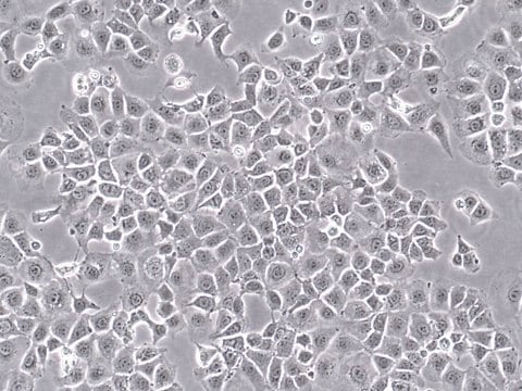
SCC178
AT-3 Mouse Mammary Carcinoma Cell Line
Mouse
Synonym(s):
Ms mammary gland cells
Sign Into View Organizational & Contract Pricing
All Photos(3)
About This Item
UNSPSC Code:
41106514
eCl@ss:
32011203
NACRES:
NA.81
Recommended Products
product name
AT-3 Mouse Mammary Carcinoma Cell Line, AT-3 mouse mammary tumor cell line may be used to develop mouse models for mammary cancer studies.
biological source
mouse
technique(s)
cell culture | mammalian: suitable
shipped in
ambient
General description
The AT-3 cell line was isolated from an autologous mammary tumor from cells originating in a transgenic MTAG mouse model . Mammary tumors arising in MTAG mice closely mimic changes observed in human breast carcinomas over the course of disease progression . AT-3 cells are a useful mouse model for studies of tumor physiology that are representative of typical disease states in humans.
Source:
The AT-3 cell line was derived from primary mammary tumor cells of a transgenic MTAG mouse. The MTAG mouse model expresses the polyoma virus middle T antigen under control of the MMTV-LTR promoter .
Note: AT-3 cells demonstrate diverse morphologies in culture.
Source:
The AT-3 cell line was derived from primary mammary tumor cells of a transgenic MTAG mouse. The MTAG mouse model expresses the polyoma virus middle T antigen under control of the MMTV-LTR promoter .
Note: AT-3 cells demonstrate diverse morphologies in culture.
Tumor-induced immunosuppression is a common phenomenon in cancer and an important consideration for cancer immunotherapy. However, most studies on tumor-induced immunosuppression rely on tumor implant models, in which the disease progresses rapidly and usually causes non-physiological host responses that do not recapitulate normal tumor development and progression . Models that generate autochthonous tumors are therefore valuable for providing more physiologically-relevant systems for investigating tumor-host interactions and longer-term immunosuppressive effects.
Cell Line Description
Cancer Cells
Application
AT-3 mouse mammary tumor cell line may be used to develop mouse models for mammary cancer studies.
Research Category
Cancer
Oncology
Cancer
Oncology
This product is sold solely for research use per the terms of the “Restricted Use Agreement” which govern its use as detailed in the product Data Sheet or Certificate of Analysis. For information regarding any other use, please contact licensing@emdmillipore.com.
Quality
• Each vial contains ≥ 1X10⁶ viable cells.
• Cells are tested negative for infectious diseases by a Mouse Essential CLEAR panel by Charles River Animal Diagnostic Services.
• Cells are verified to be of mouse origin and negative for inter-species contamination from rat, chinese hamster, Golden Syrian hamster, human and non-human primate (NHP) as assessed by a Contamination CLEAR panel by Charles River Animal Diagnostic Services.
• Cells are negative for mycoplasma contamination
Storage and Stability
Store in liquid nitrogen. The cells can be cultured for at least 10 passages after initial thawing without significantly affecting the cell marker expression and functionality.
Disclaimer
This product contains genetically modified organisms (GMO). Within the EU GMOs are regulated by Directives 2001/18/EC and 2009/41/EC of the European Parliament and of the Council and their national implementation in the member States respectively. This legislation obliges {HCompany} to request certain information about you and the establishment where the GMOs are being handled. Click here for Enduser Declaration (EUD) Form.
Unless otherwise stated in our catalog or other company documentation accompanying the product(s), our products are intended for research use only and are not to be used for any other purpose, which includes but is not limited to, unauthorized commercial uses, in vitro diagnostic uses, ex vivo or in vivo therapeutic uses or any type of consumption or application to humans or animals.
Unless otherwise stated in our catalog or other company documentation accompanying the product(s), our products are intended for research use only and are not to be used for any other purpose, which includes but is not limited to, unauthorized commercial uses, in vitro diagnostic uses, ex vivo or in vivo therapeutic uses or any type of consumption or application to humans or animals.
Storage Class Code
10 - Combustible liquids
WGK
WGK 2
Certificates of Analysis (COA)
Search for Certificates of Analysis (COA) by entering the products Lot/Batch Number. Lot and Batch Numbers can be found on a product’s label following the words ‘Lot’ or ‘Batch’.
Already Own This Product?
Find documentation for the products that you have recently purchased in the Document Library.
Trina J Stewart et al.
Journal of immunology (Baltimore, Md. : 1950), 179(5), 2851-2859 (2007-08-22)
Ag-specific and generalized forms of immunosuppression have been documented in animal tumor models. However, much of our knowledge on tumor-induced immunosuppression was acquired using tumor implant models, which do not reiterate the protracted nature of host-tumor interactions. Therefore, a transgenic
C T Guy et al.
Molecular and cellular biology, 12(3), 954-961 (1992-03-01)
The effect of mammary gland-specific expression of the polyomavirus middle T antigen was examined by establishing lines of transgenic mice that carry the middle T oncogene under the transcriptional control of the mouse mammary tumor virus promoter/enhancer. By contrast to
Multiphoton Microscopy of FITC-labelled Fusobacterium nucleatum in a Mouse in vivo Model of Breast Cancer.
Parhi, et al.
Bio-protocol, 13, e4635-e4635 (2023)
Weijie Zhang et al.
Cell, 184(9), 2471-2486 (2021-04-21)
Metastasis has been considered as the terminal step of tumor progression. However, recent genomic studies suggest that many metastases are initiated by further spread of other metastases. Nevertheless, the corresponding pre-clinical models are lacking, and underlying mechanisms are elusive. Using
C Patrick Reynolds et al.
Methods in molecular medicine, 111, 335-350 (2005-05-25)
Rodent models provide an important means of assessing antitumor activity vs toxicity for new cancer therapies. Tumors are often grown subcutaneously on the flank or back of animals, allowing accurate serial determination of tumor volume with calipers by measuring the
Our team of scientists has experience in all areas of research including Life Science, Material Science, Chemical Synthesis, Chromatography, Analytical and many others.
Contact Technical Service


