Accéder au contenu
Merck
Toutes les photos(1)

Key Documents

C1956

Sigma-Aldrich

Monoclonal Anti-Calcineurin (α-Subunit) antibody produced in mouse

clone CN-A1, ascites fluid

Se connecterpour consulter vos tarifs contractuels et ceux de votre entreprise/organisme


About This Item

Numéro MDL:
Code UNSPSC :
12352203
Nomenclature NACRES :
NA.44

Source biologique

mouse

Niveau de qualité

Conjugué

unconjugated

Forme d'anticorps

ascites fluid

Type de produit anticorps

primary antibodies

Clone

CN-A1, monoclonal

Contient

15 mM sodium azide

Espèces réactives

human, bovine, rat

Technique(s)

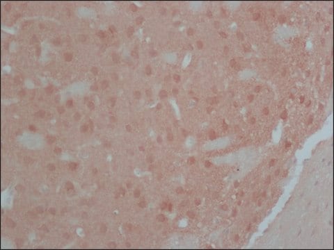
immunohistochemistry (formalin-fixed, paraffin-embedded sections): suitable using neurons in human ganglia
indirect ELISA: suitable
microarray: suitable
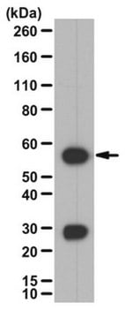
western blot: 1:10,000 using rat brain extract

Isotype

IgG1

Numéro d'accès UniProt

Conditions d'expédition

dry ice

Température de stockage

−20°C

Modification post-traductionnelle de la cible

unmodified

Informations sur le gène

human ... PPP3R1(5534)
rat ... Ppp3r1(29748)

Description générale

Calcineurin (CaN)(heterodimeric enzyme) is a Ca2+/calmodulin (CaM)-stimulated protein phosphatase 3 (PPP3) consisting of a catalytic A subunit (CnA) and a Ca2+-binding regulatory B subunit (CnB). The calcium-binding regulatory subunit (calcineurin B) is coded by the PPP3R1 gene located on human chromosome 2. Protein phosphatase 3, regulatory subunit B, α (PPP3R1) is also known as CALN, CCN1, CNA1, CALNA, PPP2B and CALNA1. The regulatory subunit calcineurin B is widely expressed in brain while the catalytic subunit (calcineurin A) is abundantly expressed in Hassall′s corpuscles, localized in the thymic medulla and represents the terminal stages of thymic medullary epithelium.
Monoclonal Anti-Calcineurin (α-subunit) (mouse IgG1 isotype) is derived from the CN-A1 hybridoma produced by the fusion of mouse myeloma cells and splenocytes from immunized BALB/c mice.

Spécificité

In immunoblotting, the product recognizes an epitope located on the α-subunit of calcineurin (61 kDa, also called calcineurin A) and does not cross-react with the β-subunit.

Immunogène

bovine brain calcineurin

Application

Monoclonal Anti-Calcineurin (α-Subunit) antibody produced in mouse has been used in:enzyme linked immuno sorbent assay (ELISA) ,immunohistochemistry, western blotting

Actions biochimiques/physiologiques

Calcineurin is a major soluble calmodulin-binding protein in the brain. It stimulates the expression of the surface molecules CD83, CD80, CD86, CD40 and HLA-DR by promoting secretion of inflammatory cytokines IL-6, TNF-α and IL-1β by human PBMC-derived dendritic cells. It plays an important role in signal transduction, activation of T cell. Calcineurin is an excellent marker enzyme for the detection of neuronal activity and synaptic plasticity after brain damage.

Clause de non-responsabilité

Unless otherwise stated in our catalog or other company documentation accompanying the product(s), our products are intended for research use only and are not to be used for any other purpose, which includes but is not limited to, unauthorized commercial uses, in vitro diagnostic uses, ex vivo or in vivo therapeutic uses or any type of consumption or application to humans or animals.

Vous ne trouvez pas le bon produit ?  

Essayez notre Outil de sélection de produits.

Code de la classe de stockage

10 - Combustible liquids

Classe de danger pour l'eau (WGK)

nwg

Point d'éclair (°F)

Not applicable

Point d'éclair (°C)

Not applicable


Certificats d'analyse (COA)

Recherchez un Certificats d'analyse (COA) en saisissant le numéro de lot du produit. Les numéros de lot figurent sur l'étiquette du produit après les mots "Lot" ou "Batch".

Déjà en possession de ce produit ?

Retrouvez la documentation relative aux produits que vous avez récemment achetés dans la Bibliothèque de documents.

Consulter la Bibliothèque de documents

Colin Rickman et al.
The Journal of biological chemistry, 279(13), 12574-12579 (2004-01-08)
Synaptotagmins are membrane proteins that possess tandem C2 domains and play an important role in regulated membrane fusion in metazoan organisms. Here we show that both synaptotagmins I and II, the two major neuronal isoforms, can interact with the syntaxin/synaptosomal-associated
H Bito et al.
Cell, 87(7), 1203-1214 (1996-12-27)
While changes in gene expression are critical for many brain functions, including long-term memory, little is known about the cellular processes that mediate stimulus-transcription coupling at central synapses. In studying the signaling pathways by which synaptic inputs control the phosphorylation
Anindit Mukherjee et al.
American journal of physiology. Renal physiology, 320(5), F719-F733 (2021-03-16)
Phosphorylation of the thiazide-sensitive NaCl cotransporter (NCC) in the distal convoluted tubule (DCT) is altered rapidly in response to changes in extracellular K+ concentration ([K+]). High extracellular [K+] is believed to activate specific phosphatases to dephosphorylate NCC, thereby reducing its
Jolene M Kim et al.
Neurochemical research (2018-01-05)
Impaired interactions between Calcineurin (Cn) and (Cu/Zn) superoxide dismutase (SOD1) are suspected to be responsible for the formation of hyperphosphorylated protein aggregation in amyotrophic lateral sclerosis (ALS). Serine (Ser)- enriched phosphorylated TDP-43 protein aggregation appears in the spinal cord of
Impaired CuZn Superoxide Dismutase (SOD1) and Calcineurin (Cn) Interaction in ALS: A Presumed Consequence for TDP-43 and Zinc Aggregation in Tg SOD1G93A Rodent Spinal Cord Tissue
Kim Jm, et al.
Neurochemical Research, 44(1), 228-233 (2019)

Notre équipe de scientifiques dispose d'une expérience dans tous les secteurs de la recherche, notamment en sciences de la vie, science des matériaux, synthèse chimique, chromatographie, analyse et dans de nombreux autres domaines..

Contacter notre Service technique